Team:HSiTAIWAN/Notebook/FA/Pb

Oh hello there,I am Charlie,a trusty member of the "Herb Tasters" and also the brainiest E. coli in the colony.
I know all the secrets of Chinese herbs and their magical healing powers.
If you are up to a challenge,find me at team HSiTW at the jamboree.
I am the one in a straw hat,showing them pearls.I will be waiting.
Muhahahahaha
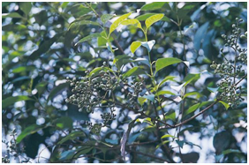
Hi there! My name is Nu Zhen Chi. This is how I look like.
Take a closer look; guess which part of me is used as medicine?
(1) the root 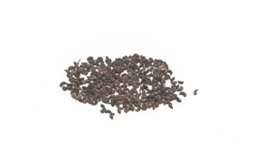
(2) the stem
(3) the leaf
(4) the seed
Ans.(4) the seed
Name: 女貞子 (Nu Zhen Chi)
Botanical Name: Ligustrum lucidum Aiton
I can treat people who are yin deficient, and liver problems that cause dizziness,cataract of the eyes,
lower back pain, premature graying of the hair and tinnitus.
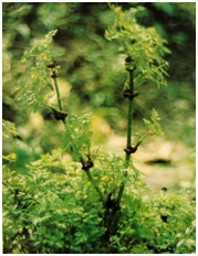
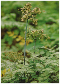
Hello! My name is Chuan Xiong. This is how I look like.
Make a guess, which part of me is used as medicine?
(1) the root 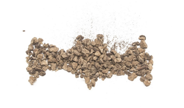
(2) the stem
(3) the leaf
(4) the seed
Ans.(1) the root
Name: 川芎 (Chuan Xiong)
Botanical Name: Ligusticum chuanxiong Hort
I help with blood regulation to prevent relevant to blood stasis and non-stop bleeding.I can also strengthen your qi circulation.
In addition, I relieve you of physical pain,
such as headaches, abdominal aches, chest pain, and muscle pain.
Finally, I free the ladies of menstrual disorders and amenorrhea.
What’s up? My name is Dang Gui. I can:
(1) stop coughing 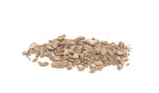
(2) regulate mense
(3) reduce internal heat
Ans.(2) Regulate mense
Name: 當歸 (Dang Gui)
Botanical Name: Angelica sinensis (Oliv.) Diels
I can remove blood stasis and clots, so I am usually used to regulate menses,lubricate intestines to correct constipation, reduce swelling, expel pus.
Reference
臧堃堂 (2005) 中華材輕百科-現代版本草綱目,山岳文化出版社,台北
Non-Profit Organization Brion Research Institute of Taiwan.
Chinese Herb Gallery. Jade Institute
Herbal Glossary. Shen-Nong- Chinese Traditional Medicine
Acknowledgement
Thank you for Non-Profit Organization Brion Resaerch Institute of Taiwan that provide us Chinese herbs and photos.
Functional Array
- Pb
- Cu
- As
- CYP
Chinese Medicine testing
- Pb
- Cu
- As
- CYP
Functional Array Pb
2015/11/12
Topic:Bacteria Culturing
Material:
old agar with E coli
coating bar 2
new agar plate 2
chloramphenicol
curette 2
Procedure:
1.Gather bacteria from old agar plate with curette
2.Put bacteria in shaking incubator for 15 minutes
3.Add more chloramphenicol and bacteria to one of thenew agar plate and add only bacteria to the other plate
4.Spread the bacteria evenly with coating bar
5.But new agar plates in incubator
Results: are expectednext week
The number of reconstructedbacteriawas far less then we had expected. The previous agar plate showed abnormal colonies, and the DNA density results ran out low. We assume the antibiotic wearing off over time, or the cloning vectors unable to successfully be inserted. Therefore, we tried adding more antibiotics to solve the solution.
2015/11/19
Topic: PCR & agar making
Material:
10X PCR buffer *10μl
50mM MgCl2 *3μl
10mM dNTP *2μl
5U/μlDNA Polymerase *0.4μl
dd2 *74.6μl
10μM forward primer *5μl
10μM reverse primer *5μl
Blue dye *2μl
10g/500 ml LB *10ml
LB *4g
Agar bacteriological *3g
dd2 *200ml
Procedure:
1.Mix the bacteria that have been incubated since last week with PCR buffer, dNTP, DNA polymerase, MgCl2, dd2, and primers into a tube.
2.Separate the mixture into six smaller tubes and place them into the thermal cycler.
3.Prepare the gel electrophoresis. Build theplate, and thenheat the agar roll.
4.Add red dye to the agar roll and spread them across the built module. Reduce the bubbles in the agar roll.
5.Retrieve the six tubes after PCR, mix them with the blue dye, and add them in individual wells.
6.Turn on the electricity for (120V, 400mA) 35 minutes.
7.Scan the gel under infrared ray and select the RNAwe are interested inexamining.
8.Target the colonies that are consistent with the gel RNA. Culture them in LB for two tubes(5ml each).
1.Add LB, agar bacteriological, and dd2 into one single tube.
Results:
We found two colonies of bacterium that matchthetargeted RNA. Accordingly, we are planning to culturethem and check ifthey may convey the right plasmid.

2015/11/26
Topic: Agarose Gel Extraction
Material:
nsert*800ng
Vector*500ng
10XBuffer (CutSmart)*2
10X BSA
Spe1*3.0μl
Xba1*1.5μl
dd2*52.8μl
1/10000 gel green
Agarose*40ml
Dye*12μl
PE buffer *0.75ml
3μl/1mg QG buffer
1μl/1mg isoprophanol
DNA ligase
Procedure:
1.Add the restriction enzymes, buffer, and BSA into two tubes. One tube containsthe insert, and another containsthe vector.Adddd2 and make up to 40μlin both tubes.
2.Incubate both tubes at 37°C for 1 hour.
3.Add 6μl of dye into each tube. Prepare the agarose with gel green dye.
4.Run the gel electrophoresis at 120V, 400mA for 30 minutes.
5.Slice the glowing DNA under UV light and remove excess agarose.
6.Put the DNA slices in the clean tubes. Add QR buffer and heat the tubes at 23°C until the gel slices are completely dissolved.
7.After the gel slices are liquified, add isoprophanol.
8.Centrifuge it for three times. Firstly, centrifuge it and discard flow-through agarose.Then, centrifuge it again and add 0.75ml PE buffer.Finally, centrifugeitagain and discard the residual ethanol.
9.Mix 10X buffer, DNA ligase, vector*4μl, and insert*13μlaltogether. Place the mixturein a 16°C water incubator for ligation.
Results: are expectednext week
The number of reconstructedbacteriawas far less then we had expected. The previous agar plate showed abnormal colonies, and the DNA density results ran out low. We assume the antibiotic wearing off over time, or the cloning vectors unable to successfully be inserted. Therefore, we tried adding more antibiotics to solve the solution.
2015/11/27
Topic: Transformation
Material:
ligated plasmid*10μl
DH5αbacterium*100μl
LB broth(without antibody) *900μl
Agar plates*2
Procedure:
1.Add the ligated plasmid to the tube of DH5αbacterium. Then put the mixture onice for 30 minutes.
2.Heat shock the tube of mixturefor 60 second ina warm water bath at 42∘C. Then, place itonice for 3minutes.
3.Add the 900μl of LB broth into the tube.
4.Put thetubein a shaking incubator for 1 hour.
5.Centrifugefor 2 minutes at 6000rpmand remove 200μl of excess broth on the top.
6.Divide the mixture into 50μland 150μlrespectively, and spread evenly across two agar plates.
2015/12/10
Topic: PCR of Final Plasmid
Material:
10X Bfr*35μl
50mM MgCl2*10.5μl
10mM dNTP*7μl
5U/μl DNA Polymerase*1.4μl
dd2*261μl
10μM forward primer(I721002)*17.5μl
10μM reverse primer(B0015)*17.5μl
Agarose*1.5g
0.5X TAE*98.5g
1/20000 red dye*2μl
Marker*4μl
Procedure:
1.Pick out white colonies from the agar plate.
2.Run PCR.
Results:
98% of the E coli on the agar plate wecultivated last time has shown a red fluorescent. Supposedly, a successfully implantedE coli should not have expressedthis characteristic unless they react with lead ions. Accordingly, weassumed that the backbone stucktogether without containing the insert genes. Thus, we picked out the colonies that werepale white. After PCR, our results showedno signs of successful plasmid.
Results: None
2015/12/17
Topic: PCR, Non-gel Extraction, Ligation
Material:
10X Bfr*20μl
50mM MgCl2*6μl
10mM dNTP*4μl
5U/μl DNA Polymerase*0.8μl
dd2*149.2μl
10μM forward primer(I721002)*10μl
10μM reverse primer(B0015)*10μl
Agarose*1.5g
0.5X TAE*98.5g
1/20000 red dye*2μl
Marker*4μl
Positive control(B00321~I721001)
Vector fragment(backbonePSB1C3)*18μl
10X cutsmart*2μl
CIP(calf intestinalphosphatase)*0.5μl
EB bfr*40μl
QG bfr*300μl
DNA ligase*1μl
10X bfr*2μl
Insert(B00321~I721001)*12μl
dd2*1μl
Procedure:
1.Mix the vector fragments(backbone)with CIP and place in the water incubator for 30 minutes at 37degrees.
2.Extract the sample to get rid ofCIP.Because there is no gel in the sample, so no need to remove the gel.
3.Ligase the sample with the interested part(B00321~I721001).
4.Pick out the white colonies from the cultural plates, from previous agar platesthat were cultured on 11/26and 12/10respectively.
5.Run PCR of the white colonies.
Results:
Last time, we didn’t find any strips of DNA that matchedour interested DNA. Today, however, several colonies that we cultured last time turn out to be white,which was a possible feature of a successful E. coli. So we run a PCR to test whether the white E. coli has been successfully implanted. The results werenegative. We still don’t have a completed cell. We thought that the binding of the backbone’s two ends might affect the results. Thus, we decided to knock the sticky ends out and try to ligase the plasmid again.

Interested DNA lenth:170bp
Results: none
2015/12/24
Topic: Plasmid Mini, DNA Restriction, Gel Electrophoresis
Material:
PS1 Buffer
Ethanol*100ml
RNase A
W2 Buffer
50% Glycerol
PS2 Buffer*200μl
PS3 Buffer*300μl
Wash Buffer*1000μl
dd2*50μl
DNA(final plasmid)*10μl
10X Buffer(cutsmart)*10μl
10X BSA*10μl
Restriction enzyme(Spe1)*2.5μl
Restriction enzyme(Hpa1)*2.5μl
dd2*65μl
Agarose*1.5g
0.5X TAE*98.5g
1/20000 red dye*2μl
Marker*4μl
Procedure:
1.Extract the DNA from the six chosen colonies with the plasmid mini kit. Collect them in six individual tubes.
2.Cut DNA at the restriction site of Spe1~Hpa1(approximately 100bp).
3.Measure the concentration of DNA from each tube. Eliminate the sample with a low concentration(17.7ng/μl).
4.Run the remaining DNA samplesin a gel electrophoresis.
5.Searchfor DNA parts at the length of 100bp.
Results:
Our white E. coli colonies could have two possible results. First, the plasmid might have a reversed insert site. In this case, there should be a DNA length about 500bp when we run the infrared sensor. Second, theplasmid has been successfully inserted.If this is the case, we should see a DNA length about 100bp.
Our results came out with no DNA length near 100. However, we found four plasmids showing a sequence of DNA with the same length, which we assume itis the backbone. Three ofthe plasmids show nothing else;while one plasmid shows a DNA with the length around 500bp. The last plasmid is believed to be the one with a wrong insert.Because we did not find the DNA, we hypothesized that the three plasmids have a different DNA sequence than the last one. In other words, we think that the three plasmids convey the correct DNA. Yet, the infrared sensor device did not show any DNA we were looking for. So, we presumed that the enzyme(Hpa1) could be mild, causing poor visibility of the DNA. Next time, we plan to enhance the quantity of the enzyme. Also, we will increase the density of gel so that the DNA could be more concentrated.

The plasmid to the right shows an additional DNA.
The four plasmids on the right show a same DNA part.
Interested DNA:100bp
Results: none
2015/12/31
Topic: Gel Electrophoresis
Material:
DNA(sample1 DNA density: 107.9ng/μl)*3μl
DNA(sample3 DNA density: 413.4ng/μl)*0.8μl
DNA(sample5 DNA density: 821.4ng/μl)*0.4μl
10X BSA*6μl
10X Bfr*6μl
Spe1*2.4μl
Hpa1*2.4μl
dd2*39μl
Green dye*2μl
1.5% Agarose*25μl
100bp Marker*2μl
Procedure:
1.Choose three colonies that differ the most. Measure the density of DNA in each.
2.Aquire thecorrect amountof samples to make the equal amount of DNA.
3.Run the gel electrophoresis.
Results
Although we have added more enzymes, there is still no sign of DNA around 100bp. The result is similar to that from last time, but with a clearer view of the DNA. Due to the fact that features of the colonies we have chosen accord to a successfully inserted colony.There should be a DNA strain around 100bp, unless, the sequence of our E coli. contradicts logically. We will check whether there is any problem in our sequence before the RFP.

Interested DNA:100bp
Results: none
2016/01/14
We now face difficulties of getting the correct DNA length. Thus, we ran a primer on two different plasmids, from the RFP site to the Lead Promoter site. After matching the DNA sequence, we couldn’ t find the Lead Promoter site. However,we found the DNA sequence of B0032 at 471~483bp, the sequence of I721002 at 491~889bp and an incomplete part of B0015 at 897~. This result (figure 1)fits perfectly if we consider the whole insert to be reversed. The reason for the missing sequence of the Lead Promoter may be because the primer couldn’t go any further. So, we took a closer look at the three DNA sequences we matched. The B0032 sequence was negative(reversed), but the other two were positive(not reversed). To shorten up, the B0032 has been reversed, and the whole insert has also been reversed in the backbone. Next time, we will reinsert the insert partsand make sure we have the right plasmid (figure 2)through quick PCR.

2016/01/25
Topic: Gene Extraction, Ligation
Material:
CIP*1μl
restriction enzymes(Spe1)*5μl
restriction enzymes(Xba1)*2.5μl
10X cutsmart*2μl
1/20000red dye*2μl
blue dye
Marker*4μl
Agarose*1.5g
0.5X TAE*98.5g
Vector(backbone)*5μl
Insert(B0032~I721001)*12μl
3μl/mg QGbfr*630μl
DEbfr *750μl
dd2*80μl
10X bfr*2μ
Procedure:
1.Redo the ligation process and then extract the insert and vector from individual plasmids.
Results:
None. Results areexpected after transformation and PCR.
2016/01/26
Topic: Transformation
Material:
Ligated plasmid*10μl
DH5αbacterium*100μl
LB broth*900μl
Agar petri dish*1
Procedure:
1.Transform the plasmid into the DH5α
2.Culture the bacterium.
Results:
*The amount of bacterium shown after centrifugation was unusuallylittle.
Results are expected after PCR.
2016/01/27
Topic: PCR of final plasmid
Material:
10X bfr*(35+35)μl
50mM MgCl2*(10.5+0)μl
dNTP*(7+7)μl
F primer(E1010)*(17.5+7)μl
R primer(B0032)*(17.5+7)μl
Polymerase*(1.4+4.2)μl
dd2*(261.1+289.8)μl
0.8X TAE*140ml
Agarose
1/1000 AMP*8μl
LB broth*8ml
*We tried a new buffer that included MgCl2, so we didn’t add in any MgCl2 the second time we did PCR
Procedure:
1.Pick out 62 colonies from the former petri dish.
2.Do PCR.
3.Pick out 2from the 9 colonies that have our interested DNA length.
4.Culture the 2 colonies in LB broth.
5.Send the samples for sequencing.
Results:
Every colony on the agar plateappears to be white, which is what a correctly ligased E. coli should express. To increase the chance of succeedingin finding the correct colonies, we picked 62 colonies for PCR. 9(1-2, 1-3, 1-4, 1-5, 1-6, )out of the 62 tend to have the rightlength of DNA(about 700bp). And to make sure that the sequences are accurate, we chose 2 colonies(1-6, 2-18)which are most valid after infrared lightscanning. We will send the samples in for further sequencing.

2016/02/01
After sequencing, we confirmed that the DNA is indeed what we need. To testthe E. coli accessibility, we are going to performethree experiments,including howthe E.coli react to Pb in different concentrations([Pb]), what factors may alterthe expression of the E. coli, and the survival ratesof E. coli in different living conditions. We makea table that lists four variables(restriction of growth, E. coli amount, time,and[Pb]) to record different results. It was our original plan (Figure1and Table 1).
In order to assure that there are equal amountsof bacteriain the tubes,we cultured the E. coli in a single tube, andthenevenly divided into all tubes. Next, we added5differentconcentrations of Pb into the 5tubes. However, at this point, the professor reminded us to be aware of the Pb ions penetration-time. The different concentration of Pb might affect thebacteria’s absorption speed, thus influence the amount of Pb in the cells. To control these potential biases, he advisedus to observe Pb in different concentrations, and control three variables in terms of restriction of growth, time duration, and amount of E coli. We then changed our plan and performed the new strategies.
First, we prepare 10 tubesthat contain different [Pb]concentration levels, andevenly add the E. coli in each tube(Figure2and Table 2). Then, culture the tubes for one nightto ensure all bacteriahaveabsorbedthe maximum amount of Pb ion. Then, we will observe the remaining E. coli (some might die in high concentration of Pb) and examine the differencesbetween one another via O.D. Finally, we will choose the most suitable Pb concentration as the goldstandard to conduct the remaining experiments. By completing these processes, we can both figure out the best situation to collect our results and test the survival rate of our E. coli.

Topic: Preparing E. coli
Material:
Ampicillin*6μl
LB broth (without antibiotics)*6ml
E. coli (cloned)*3μl
E. coli as vector (J23102)*3μl
Tube*2
Procedure:
1.Mix two tubes of LB broth with Ampicillin.
2.Add the cloned E. coli and E. coli(J23102)into 2 individual tubes, respectively.
2016/02/02
Topic: Functional Array
Material:
E. coli (cloned)*45μl
E. coli as vector(J23102)*5μl
LB broth (without antibiotics)*10000μl
Ampicillin*10μl
104ppm Pb (aq)*(100+100)μl
ddH2O*9000μl
Cultivation tube*10
Dilution tube*9
Procedure:
1.Dilute the Pb aqueous solution by adding ddH2O to make 9different levels of concentration,respectively, 104(ppm), 103, ..., 10-4.
2.Prepare LB broth by adding ampicillin.
3.Make 9 cultrivation tubes that contain 9 differentlevels of Pb concentration.[formula: 100μl of Pb(aq),5μl of bacterium and 900μl LB to form 1c.c of fresh mixture]
*Because the aqueous solution has been diluted again in the mixture, the true concentration inthe tubes should drop one foldof ten.
4.Cultivate the 10 tubes(inclulding one control tube)in a shaking incubatorovernight.
Results:
Results are expected the next day.
2016/02/03
Topic: Functional Array2
Material:
Pb(NO3)2*5g
ddH2O*45ml
LB broth*4500μl
Ampicillin*4.5μl
E. coli (cloned)*(200+20)μl
E. coli as vector (J23102)*5μl
Petri dish*2
Coating bar*2
Procedure:
1.Run an O.D test on the tubes from last time.
2.Check the E. coli under UV light.
3.Make a fresh tube of Pb aqueoussolutionin the concentration of 105ppm.
4.Dilute the solution into 8 different levels.(105, 104, ..., 10-2ppm)
5.Spread 100μl of E. coli per fresh agar dish. Add 4 different Pb solution on 4 corners of the plate. (Do not shake the petri dishes!)
6.Make 4 cultivation tubes like last time with the concentration of 10, 103, 102, 101ppm and a tube for reference without Pb. (formula of 1c.c. mixture: LB broth*900μl, Amp*0.9μl, bacterium*5μl, Pb(aq)*100μl)
Results:
OD results

According to the results in the table above, the concentration of Pb between 101to 10-7ppm doesn’t affect the survival of E. colimuch. There isnosignificant finding in this experiment.
Compare to the control tube, the E. coli in the experimental tubes didn’t express any obvious glow(Figure 1 and 2). There were two Victor Shen 2016/2/3possibilities. First, the experiment indeed failed. Second, the E. coli actually did react, but was just too faint for our naked eyes to observe. Surprisingly, our bacterium did express a hazy fluorescent(Figure 3).So, we assumed the process is accessible, but the level ofPb ions might be too low. Much to our shock, we mistook the former [Pb] 102folds higher. Every solution was 102lower than the what we mean to prepare.
Meanwhile, we concerned that LB broth might somehow react with the Pb solution, and then alter the proper functionof the E. coli. Hence, we tried anothermethod to test the results of bacteriain different [Pb] levels.We first spread our E. coli onto the agar plates to make sure they have the necessary survival requirements. Next, we dripped small amounts of Pb solution onto the surface of our E. coli. By doing this, we expect no other materials from the broth could interfere the reaction.

2016/02/18
Topic: Fluorescence Spectrometry
Material:
LB broth*1750μl
Control bacteria(J61002)*250μl
100μl/well Old bacterial fluid in different levels of [Pb]*100*14μl
Thermo Varioskan Flash
Procedure:
1.Mix the bacteria clusters at the bottom of the Eppendorf tubes.
2.Dilute the control bacteria 2 times into 7 tubes. (1, 2-1, 2-2, 2-3, 2-4, 2-5, 2-6)
3.Add the control and bacterial fluid into the wells of a photographic plate.
4.Place the plate into the fluorescence spectrometry machine. (excitation 585, emission 610)
Results:
Table 1 shows that the absorption levels and concentration levels increase at a direct proportion.Figure 1 shows that the spectrometer is precise and the relation between the absorption level and the concentration level.As shown in Table 1 and Figure1, we can see that the detecting device is precise and the deviation mean is low. Thus, our calculations can be accounted accurate.
In Table2, any bacteria with Pb solution added shows a higher fluorescence absorption compared to that without any solution amplified. There is a slight difference between the bacteria with Pb solution added and none. But the distribution of the absorption results appears to be random.Hence, we knowthat our E. coli does express a red fluorescence when combined with Pb ions. However, there is no significant relevance between the Pb ion levels and the E. coli’s fluorescence expressions.
In conclusion, our E. coli is capable of detecting the existenceof Pb ions, but is not able to differentiate the changes in the concentration levels of the solutions.

2016/02/25
Topic: Functional Array 2
Material:
Zn(NO3)2-6H2O*500mg
NiSO4-xH2O*500mg
CuSO4-5H2O*500mg
MnSO4-H2O*500mg
Pb(NO3)2*500mg
LB broth*900*34+100μl
Bacteria*5*34μ
Procedure:
1.Prepare 5solutions that consist of 5different metal ionsrespectively(Zn2+, Ni2+, Cu2+, Mn2+, Pb2+).
2.Dilute every tube of solution into 5 different levels of concentration (10-6, 10-4, 10-2, 100, 102ppm).
3.Dilute the lead ion solution further more. Make three other tubes of solution at the concentration of 10-7, 10-8, 10-9ppm.
4.Make a mixture of bacteria and metal ions. Formula:900μl of fresh LB broth with antibiotics, 100μl of diluted ion solution, and 5μl of bacteria.
5.Culture the tube in a shaking incubator.
Results:
We wanted to examine whether our E. coli has a strong singularity and if the presence of other metal ions may influence the bacteria’s absorption ability. So, we tried out 4 othermetal ions in additional to leadions. Furthermore, we wanted to test the detecting ranges of our E. coli. According to our previous functional tests, we were able to conclude that our bacterial device was capable of detecting Pb ions less than 10-7ppm. Therefore, we will try to challenge its lowest limitthis time.
2016/02/26
Topic: Functional Array2
Material:
Bacteria as control (J23102)*250μl
LB broth used as blank*250μl
E. coli used as blank*250μl
Different solutions consisting different metal ionsand BBa_K1613017
Procedure:
1.Vortex to mix the bacteria clusters at the bottom of the Eppendorf tubes.
2.
3.Add two control sets and experimental set into the wells of a photographic plate.
4.Place the plate into the fluorescence spectrometry machine. (excitation 585, emission 610)
Results:
In this study, we used functional arrays to examine the singularity. We expected to find that E coli only responded to lead ions. But we did not get the results we expected. Table 1 shows the control samples we made. In our experiment, we made two sets of control:one was fresh LB broth and E. coli; another one was J231002 in different solutions at the concentration level of 10-3ppm. Table 2 shows the fluorescence absorption of different metal ions. The results suggest that our E coli were significantly responded to lead ions. However, our E coli were also significantly responded to other metal ions. We suspected that the bacteria havea poor singularity bond between lead ions, so they also react with other metal substances. Accordingly, we decide to expend length in time,allowing sufficient time for the E. coli to interact with lead ions, and distinguish the differences of lead from other metal ions.In the previous experiment, the reaction duration was 24hours. This time, we plan to prolong the reactionduration to 1 week.

2016/03/03
Topic: Functional Array3 (fluorescence microscope)
Results:
*Exposure Time: 350ms
*During editing, we increased the brightness of every picture 100% in order to enhance the difference between the background color.
After discussion, we suspected that living bacteria might disrupt the detecting of fluorescence protein. A fluorescence spectrometry machine doesn’t only detect the fluorescence level, but it also calculates several other factors. So, we decided to examine the bacteria through a fluorescence microscope, in that way, we could rule out other factors except the fluorescence of the bacteria.
Based on the picturesbelow, they obviously showthat the bacteria consisting of lead expressesa clear glow compared to other experiment samples.Thus, we can conclude that our E. coli dohave a specific characteristic.

2016/04/28
Topic: Preparing Toxic Solution
Material:
CuSO4-5H2O*2.0g
105ppm Pb(NO3)2*10ml
Tube*13
ddH2O*118ml
Procedure:
1.Measure 2g of Cu2+crystal and pour it into a tube. Add ddH2O to 10ml.
2.Dilute the solution by 10 times in the sequence of 105 104 103 102 101 1 10-1ppm in concentration.
3.Do the same as for lead solution.
Note:
oday, our plan was to culture E.coli in five different concentrations, ranging from 103to 10-1. Thus, I first diluted the five concentrations. However, my advisor reminded me that the ion solution would be further diluted when added into the culturing broth. Either I prepare the exact same concentration in LB broth, or, I prepare a more concentrated water solution. So, I chose the latter and prepared the solution up to 105ppm. Also, I was advised thatE. coli would stop growing after 14 hours, hence, I would have to collect my sample by then. If the bacteria still remain in the 37°C incubator, eventually the cells would all die out. Another thing I learnt was that the fluorescent proteinwould begin the process of protein degradation. (Protein should be stored at -80°C, in other words, 37°C is not a nice temperature for storing protein). Given these two conditions, I should not culture the E coli today because I have school classestomorrowand I do not have two consecutive days.Therefore, I plan to culture the E. coli over the weekend.
2016/05/05
Topic: Preparing Solution and Bacteria Culturing
Material:
Na2HAsO4·7H2O*1g
ddH2O*19ml
Petri dish(ampicillin)*1
Petri dish(Chloramphenicol)*1
Alcohol lamp(70% full)*1
Steel coating bar*1
Procedure:
1.Prepare one tube of arsenic solution inthe concentration of 105ppm.
2.Dilute the solution by tenth folds, making another solution in the concentration of 104ppm.
3.Wipe the laboratory counter with adequate alcohol. Then, light an alcohol lamp in the middle of the table to provide a temporary germ-free environment (1 square meter).
4.Burn the steel coating bar until it turns bright red for sterilization. Wait about 20 seconds for the coating bar to cool off, or else, the high temperature will destroy the germ colonies later on.
5.Using the coating bar,dab a colony from a former plate. Zigzag the germ colony on a new petri dish.
6.Burn the steel coating bar again.
7.Spread the colony on the fresh petri dish further. In that case, the colonies will be more scattered. Thus, it will be easy for us to pick colonies in the future.
8.Place the petri dishes into the incubator to culture.
2016/05/06
Topic: Plasmid functional array
Material:
Cu2+solution in the concentration of 2*105to 2*10-1ppm
Pb2+solution in the concentration of 105to 10-1ppm
E. coli (K1555000) (K1613017)
Ampicillin*10μl
Chloramphenicol*10μl
LB Broth*20ml
Culturing tube*10
Procedure:
1.Create a germ-free environment with an alcohol lamp.
2.Divide the broth into two tubes. Add the appropriate antibiotic into the broth at a ratio of 1:1000.
3.Distribute the broth into the other remaining tubes.One tube should require 2ml of broth.
4.Poke the colonies on the agar plate gently with a plastic tip. Dip the tip inside the broth.
5.Add the corresponding solution into the tubes at a ratio of 1:100.
6.Place the culturing tubes into a shaking incubator overnight.
Note:
Before we open, or close the caps of containers containing LB, be sure to burn the cap for a split second. That way, we can ensure the opening to be clean and uncontaminated.
2016/05/12
Topic: Listing of Chinese Medicine and Preparing SolutionMaterials
Material:
黃岑、甘草、知母、茯苓、酸棗仁湯、薄荷、黃耆、牡丹皮、川芎、當歸、白芍、女貞子、炙甘草、炮薑、桭子、柴胡、逍遙散、四物湯、霍香
Eppendorf*20
ddH2O
Dry bath incubator
Procedure:
1.Analyze the medicine into three categories, liquid, thick broth, or solid chunks.
2.Heat the thick broth at the temperature of 100°C for 10 minutes in a dry bath incubator. Wait until the broth liquefies.
3.Dilute the fluid by 1/10 into individual Eppendorf. The formula should be: 150μl of Chinese medicine and 1350μl of ddH2O.*Thick broth often clogs up easily. So be sure to be fast.
4.Smash the solid chunks into powder, the dainty the better.
5.Measure 15mg of powder. Add ddH2O in an Eppendorf to 1.5ml. Mix the two thoroughly.
Results:
Some of the medicine stew was too thick that it did not liquefy even after heating for 20 minutes (Figure 1). And some of the medicine chunk was too hard to break down (Figure 2). So, we decided to turn to a herbal medicine professor for advice. Then, we can figure out how to deal with this situation.

2016/05/19
Topic: Preparing solution, planning the experiments
Material:
岩精*140mg
硃砂*15mg
木香*15mg
ddH2O*17ml
Procedure:
1.Add the samples into different Eppendorf.
2.Add ddH2O to the concentration of 10mg/ml.
Future Project:
We plannedto test whether Chinese medicine would suppress the growth of the E. coli. Thus, we have decided to add Chinese medicine into the culturing environment of the bacteria. Essentially, there are three methodsof conducting this experiment. First, spread Chinese medicine across the surface of the agar plates while culturing. Second, mix Chinese medicine into the agar gel. Then, culture the bacteria as usual. Third, culture the bacteria in a culturing tube and add Chinese medicine. We chose the latestmethodfor two considerations. One the one hand, the first two methods requirecounting colonies. Onthe other hand, the last method analyzes the bacteria’s growth via optical densitywhich is more reliable. Using a more objective analyzing system, we can increase the accuracy of our experiment.
Further, we dilutedChinese medicine into 5 different levels of concentration, ranging widely. Because we intend to test the survival rate of the E. coli,the concentrationrange should be broader to save time and resources.
2016/0/
Topic: Plasmid Fluorescence, ODFunctional Array 2
Material:
LB Broth*30ml
Antibiotic(Ampicillin)*1.3μl
Antibiotic(Chloramphenicol)*1.3μl
E. coli(k1555000)(Cu2+at 10-7~102ppm)*30μl each
E. coli(k1613017)(Pb2+at 10-7~102ppm)*30μl each ddH2O*(13+1.3)*2μl
Copper ion solution(10-5, 10-3, 10-1, 101, 102ppm) *10μl each
Lead ion solution(10-5, 10-3, 10-1, 101, 102ppm)*10μl each
Procedure:
1.Prepare blank samples with LB broth, ddH2O, ion solution, and antibiotics. (Formula: 1300μl of LB and 13μl of ddH2O, 1300μl of LB and 1.3μl of ddH2O, 1300μl of LB and 1.3μl of antibiotics)
2.Dilute the cultured bacterial fluid from last night by 1/10, 1/ 50. Be sure to vortex and then centrifuge the fluid briefly before dilution, that way, the E. coli will distribute more evenly.
3.Load the corresponding fluid into the well(Figure 1, 2). (200μl per well)The well for testing optical density is transparent, while the well for testing fluorescence is black.

2016/06/02
Topic: Analysisof OD, GFP, RFP
Results:
According to the bar charts(Figure 1 & Figure2), we can see that as the same bacterial fluid gets diluted, the optical density drops.Also, from the charts, we can assume that metal ion concentration levels are not relevant to the growth of E. coli. Hence, the instrument for measuring OD is functional and accurate.
In terms of the fluorescence sample, results are not going well. Aside from the sample with thehighest concentration of copper ion (102ppm), all the other testing samples have no significant absorption difference with the negative control sample (Figure 3). What’s worse, the fluorescence absorption in RFP (biosensor for lead) appears random(Figure4). Thus, we cannot even find the slightest connection between the concentration of lead ion and the intensity of RFP, whereas the negative control sample shows a stronger fluorescence than the sample with the highest ion concentration. Based on the results, we are guessing that the instrument for detecting fluorescence absorption is not reliable. So, we have to use another device in our next experiment.

2016/06/16
Topic: Transformation
Material:
Competent cell (DH5a)*15*3μ
Plasmid (K608010, K608012, K516132)*2μl for each
SOB*200*3μl
Agar plate*3μl
Procedure:
1.Add 15μl of competent cell with 2μl of plasmid into an Eppendorf.
2.Ice the bacteria in ice for 5 minutes.
3.Heatshock the bacteria in an incubator at 37°C for 3 minutes.
4.Place the bacteria in ice to recover for 2 minutes.
5.Add 200μlof SOBinto the bacteria.
6.Put the bacteria in a shaking incubator for 4 hours.
7.Spread the bacteria on an agar plate and distribute the fluid evenly with a coating bar.
8.Place the petri dish into an incubator overnight.
2016/07/18
Topic: Culturing Frozen Bacteria
Material:
Frozen bacteria (k1613017, k1555000, J23102)*100μl
Ampicillin*50μl
Chloramphenicol*50μl
Agar plate*3
Procedure:
1.Add antibiotics in the antibiotic free agar plates.
2.Distribute the bacteria evenly on the plate with acoating bar.
2016/0/
Topic: Growth RatePretest, Fluorescence Preparation, Lead Solution Preparation, Strick Out
Material:
LB*20ml
Ampicillin*20μl
Former agar plate (k1613017)
LB*6000+490*48μl
Ampicillin*3*2μl
Former agar plate (RFP positive control, k1613017)
24well plate*1
Culturing tube*24
Pb(NO3)2*8g
ddH2O*500+0.9*10ml
Coating bar*1
Fresh agar plate*3
Former agar plate (RFP positive control, k1613017, k1555000)
Procedure:
(For growth rate pretest)
1.Pick a colony (k1613017) and add it in 20μl of Ampicillin and 20mlof LB.
2.Culture for the first 4 hours without testing optical density(OD). *Because the bacteria haven’t gone into a log phase, the growth appears to be slow. There is no use in testing OD at this time.
3.Test the optical density next every 1 hour.
(For fluorescence preparation)
1.Pick two colonies (k1613017, RFP positive control) and culture them separately in 3ml of LB and 3μl of Ampicillin.
2.Culture the two tubes in an orbiting incubator for 8 hours.
3.Add 490μl LB, 5μl of lead solution, and 3μl of bacterial fluid into a 24well plate and culturing tubes.
4.There should be six different samples in each experiment set. Negative control, positive control (RFP), 101, 10-2, 10-7, 10-8ppm concentration of lead ion.
5.Incubate the two sets (well plate, culturing tube) in a shaking incubator for 16 hours.
(For lead solution preparation)
1.Add 8g of Pb(NO3)2 into 500ml of ddH2O to create a lead ion solution in the concentration of 104ppm.
2.Dilute the solution by 10 folds, from 104to 10-6ppm. *900μl of ddH2O+100μl of previous solution.
Results:
Although the bacteria colony we took contained a large variety of E. coli, we can still take the growth rate as a reference. We wanted to examine the growth phase of our bacteria, that way, we can make sure that our following experimentswill acquire healthy and fresh bacteria, which are in the log phase. In the graph below, after culturing about 7 hours, the bacteria approached 0.6, which is the perfect condition for testing. Thus, we will design our further experiments to be based on bacteria which are cultured for 7 hours or so

2016/07/20
Topic: Functional Testing of Frozen Bacteria
Material:
Lead ion solution*2*5μl (103, 10-2, 10-5, 10-6ppm)
LB*14*500μl
Ampicillin*7μl
Procedure:
1.Culture the frozen bacteria in different concentrations of lead ion for 8 hours.
2.Prepare the blank set with LBand antibiotic.
3.Test the fluorescence intensity of the sample.
Results:

From the table above, we sought that the fluorescence of bacteria did not correspond to the concentration of lead ion, which is contrary to what we tested before. Thus, we assumed that the frozen bacteria might somehow be contaminated or degraded. But, I guessed that there might be something wrong with the construct of the plasmid, or maybe the test results we did before was simply a coincidence. So, I would try to recheck the sequence that we ran before to check whether the frames have shifted. For now, we decided to re-transform the plasmid into a fresh group of E. coli, and hope to test the function again.
2016/07/25
Topic: Mini Extraction, Gel Electrophoresis, Liquid Culture, Making TAE Buffer
Material:
Mini Extraction Kit*1
0.8% Agarose*49ml
50X TAE Buffer*1ml
ddH2O
Blue Dye*12μl
EDBT
LB*4ml
Ampicillin*4μl
Procedure:
(For mini extraction)
1.Add 1.5ml of bacteria into an Eppendorf.
2.Centrifuge the sample at 12000rcf for 1 minute.
3.Remove excess fluidfrom above the tube.
4.Add 200μl of MX1 buffer onto the pellet. Vigorously vortex the sample.
5.Add 250μl of MX2 buffer into the mixture. Shake gently by rotating your hands.
6.Place the sample for 2~5 minutes at room temperature.
7.Add 350μl of MX3 buffer. Shake the sample immediately, there will appear to be a white cloudy suspension floating above the tube.
8.Centrifuge the sample at 10000rcf for 5~10 minutes.
9.Drip thefluid through a mini Plustmcolumn carefully. Do not damage the film between. Discard the pallet beneath.
10.Centrifuge the column at 7000rcf for approximately 1 minute.
11.Remove excess fluid inside the tube below.
12.Drip 500μl of WN buffer through the film.
13.Centrifuge the sample at 7000rcf for approximately 1 minute.
14.Remove excess fluid inside the tube below.
15.Drip 700μlof WS buffer through the film.
16.Centrifuge the sample at 7000rcf for approximately 1 minute.
17.Remove excess fluid inside the tube below.
18.Centrifuge again at 10000rcf for 3 minutes to get rid of remaining ethanol in the pallet.
19.Change the tube of the column into an Eppendorf.
20.Add 50μlof Elution buffer.
21.Wait for 3 minutes.
22.Centrifuge the sample at 10000rcf for 3~5 minutes. The remaining fluid will be the extraction of DNA plasmid.
23.Either use the plasmid instantly or freeze the plasmid inside a 4°C freezer.
Results:
We ran a gel electrophoresis to check whether we have successfully transformed our interested gene into our bacteria. So, we usedthe plasmid we transformed to serve as a reference to the extracted plasmid we got. The results are as the followingfigure 1. Although there is a long smear in the original plasmid, while our extracted plasmid seems clean-cut; the numbers of base pair are significantly near and can be assumed to be a successful transformation. However, since the plasmid was used 4 to 5 months ago, we cannot be sure if the plasmid has mutated or shifted frames. Thus, we decide to culture a colony of successfully transformed bacteria, and test its function tomorrow. If the results do not run out well, we will then try western blot to see if PbrR protein was ideally transcript. If that doesn’t run well either, we might choose to reconstruct our DNA.

2016/07/26
Topic: Fluorescence Spectrometry
Material:
LB (for blank)
Ampicillin (for blank)
Bacteria (k1613017, J23102)
Infinite M200 Pro (TECAN) fluorescence spectrometry
Procedure:
1.Create a sterilized environment by lighting fire from an alcohol lamp.
2.Prepare a series of Blank samples.
3.Test the optical density and fluorescence intensity, with an absorption wave length of 600nm; and an excitation length of 584nm and emission length of 607nm.
Results:
As in the table below, we can see that the experiment sampledid not differ with the negative control, which has no lead ion in the solution. Also, the sample will E. coli shows less fluorescence than our blank sample, containing only LB. Furthermore, the fluorescence of the bacteria did not grow strong overtime. Thus, we decided to culture a longer time, and if that does not prove to be well, we shall run a western blot to make sure that our bacteria is capable of producing PbrR protein.

2016/08/17
Protocol E.coli growth rate inpbsolution
Objective:
Material:
Transformed DH5α pcDNA (ctrl)on SOCagar plates
SOC broth
Amp (1000 X)
15 ml tube、50 ml tube
96 well plate
Eppendorf
Lead solution
Pipette(1000ul、200ul、20ul)
Procedure:
Day 1
1.Culture 4 tubes of DH5α(lead biosensor) in 15ml centrifuge tubeovernight: 2ml SOC with 2ulrpm:110)
2.Prepare 1000ppm, 100ppm, 50ppm, 20ppm, 10ppm, 5ppmlead solution by serial dilution
Day 2
1.Preparesix 15ml centrifugetube, each containing 1ml of SOC, and 2ul Amp.
2.Add 1mlofovernight culturedbacteria liquidinto each 15ml centrifuge tube,which makes all of them contain 1ml of SOC, and 2ul Amp, and 1ml bacteria liquid
3.Culture them for another two hours to make sure if they are able to grow.
4.Prepare five kinds of concentration oflead solution, 10μg/ml, 3μg/ml, 1μg/ml, 0.3μg/ml, 0.1μg/mlby serial dilution.(Which is prepared at day 1)
5.Prepare eighteen50ml centrifungal, each contains 29.1ml of SOC, 0.6ml of bacteria liquid, and 0.3ml lead solution(with five kinds of solution).

6.Examine the OD of each tube every 30 minutes
2016/08/23
Topic:Transform Pb plasmid
Material:
E. coli(DH5alfa)
Pb2+plasmid
SOC
Chloramphenicol(antibiotic)
Pipette
Eppendorf
15ml centrifuge tube
37°C incubator shaker
Agardish
Ice bucket
37°C shaker
Procedure:
1.Take out 60ul of E.coli DH5a from the -80 fridge and the plasmid in the -20 fridge
2.Thaw the E.coli DH5a in a ice bucket until it becomes transparent
3.Add 1ul plasmid liquid and 20ul DH5a liquid into a eppendorf
4.Put it in a ice bucket for 5 minutes
5.Heat shock the E.coli in 37 shaker for 3 minutes.
6.Put it in a ice bucket for 2 minutes(recovery)
7.Add 200ul SOC into each eppendorf
8.Put the eppendorfs into 37°C shaker for 4 hours.
9.Culture the baceteria in the eppendorfs into agar plates.