Difference between revisions of "Team:UGent Belgium/Filament"
| Line 64: | Line 64: | ||
<div class="center"> | <div class="center"> | ||
| − | <p><img src="https://static.igem.org/mediawiki/2016/f/fb/T--Ugent_Belgium--filament5.jpg" alt="fig5" height="339" width="211"></p> | + | <p class="center"><img src="https://static.igem.org/mediawiki/2016/f/fb/T--Ugent_Belgium--filament5.jpg" alt="fig5" height="339" width="211"></p> |
</div> | </div> | ||
<br> | <br> | ||
| Line 81: | Line 81: | ||
When the HABA is stoichiometrically displaced by biotin in the streptavidin-binding, a change in absorption is observed at 500nm wavelength, which provides the basis for a spectrophotometrically determination of accessible biotin levels (see figure 6).</p> | When the HABA is stoichiometrically displaced by biotin in the streptavidin-binding, a change in absorption is observed at 500nm wavelength, which provides the basis for a spectrophotometrically determination of accessible biotin levels (see figure 6).</p> | ||
<div class="center"> | <div class="center"> | ||
| − | <p class | + | <p class="center"><img src="https://static.igem.org/mediawiki/2016/7/72/T--Ugent_Belgium--filament7.jpg" alt="fig7" height="257" width="352"></p> |
</div> | </div> | ||
<br> | <br> | ||
Revision as of 22:21, 18 October 2016
Filament
Overview
In the filament work package, we try to produce a plastic filament that’s activated and susceptible biological appendage. The filament can subsequently be used for printing the desired 3D structure. Poly-lactate (PLA) was chosen as basic plastic carrier because of it’s biodegradable, optimal temperature, and generally easy-to-print characteristics. In order to enhance the function of the PLA, cfr. improve the condensation capacity, biological nucleation proteins will need to be fused to it. In order to get the proteins attached to the final structure, either purified or expressed on the membrane of whole cells, a moiety needs to be present in the filament that captures them, binds them en keeps them there (even while rinsing, or in flooded conditions).

General overview; 3D printed structure that's activated with biotin, will be succeptible for adhesion of proteins via biotin-avidin complexation
We’ve chosen to assess the biotin-(strept)avidin complex, since it’s a very robust and in most circumstances a fairtin-activated PLly forgiving binding. Also, thanks to the inertial properties of biotin, the adherence or impregnation to the filament can be accomplished in quite harsh environments, without damaging any structures.
There are three straight-forward approaches to biologically publicizing the biotin on the PLA filament:
1) Biotin can be actively linked to the filament or final structures
2) Biotin can be impregnated in PLA from where filament can be made which can then be used for 3D printing
3) Biotin can be coated on the filament or final structures
Since the first option is generally far to expensive to make bulk quantities of biotin-activated PLA (Salem et al., 2001; Ben-Shabat et al., 2006; Weiss et al., 2007), only option 2 and 3 will be assessed in this work package.
Method 2: Impregnation of biotin
In a first method we attempted, we tried to impregnate the biotin in the plastic. Therefore, we looked for a chemical condition in which a vast amount of PLA and biotin could be dissolved without changing or destroying their integrity. This condition was obtained by heating biotin-saturated dimethylformamide (DMF) to 130°C and dissolving PLA in it.

Figure 2: Experimental setup for the impregnation of biotin
After thoroughly mixing the dissolved PLA and biotin, the PLA (with biotin) can be crashed out of solution by adding solution to an excess of ethanol, saturated with biotin at room-temperature.
In this way we obtain biotin that would is completely captured by PLA in a nicely dispersed fashion. Surplus, it’s most likely that the biotin will remain in position and interwoven in the PLA fibers, even in circumstances of fog, dew, water,...
Next, the obtained, crashed out and dried PLA can be supplied to an extruder that will generate a filament which can subsequently be used as feed for a 3D printer.


Figure 3: left the set-up for the extruder and right the final generated filament
Method 3: Coating of biotin
The second method we tried, we made some kind of biotin-lacquer which can be painted on the filament and even on already printed structures. This lacquer was created by over-saturating a solvent with PLA and biotin, that classically dissolves PLA. After a simple trial and error essay, dichloromethane was selected as the optimal solvent, due to its fast working method of action and easy preparation.

Figure 4: By simply submerging a PLA fragment in this solution, a biotin-PLA coating will be adhered to the surface of the filament
Biological availability
Now we’ve tried to activate our filament by either impregnating it or coating it (see figure 5), it’s time to check whether these filaments show some biological availability towards biotin.

Figure 5: From left to right; PLA coated with biotin (method 3), PLA impregnated with biotin (method 2) and normal (untreated) PLA filament
In order to determine the biological activity of biotin on a solid carrier, a protocol was found based on a biological displacement reaction between the accessible biotin on the structure, and a 4-Hydroxyazobenzene-2-carboxylic acid (HABA) solution as described by Li, et al. (2007). When the HABA is stoichiometrically displaced by biotin in the streptavidin-binding, a change in absorption is observed at 500nm wavelength, which provides the basis for a spectrophotometrically determination of accessible biotin levels (see figure 6).

Figure 6: Principle for determination of biologically available biotin on and in a PLA fiber. Adapted from Li et al. (2007).
In a first step, a concentration array needs to be made to ascertain the concentration of biologically available biotin in a standard solution. The concentration we’ll get from the absorbance measurement will be compared to the amount of biotin that’s in the standard solution (after weighing, and making the solution, see figure 7). For every increment of volume (10µl of standard solution), the absorbance is measured and the concentration of biotin is determined using equation 1.
![]()
Equation 1: Determination of Biotin concentration of a solution based on an absorbance measurement.
In order to determine the availability of biotin on the fibers itself, the HABA-streptavidin solution was prepared and a pre-washed and pre-weighed pieces of PLA were iteratively added, vortexed for 1 minute and removed again. The resulting solution was measured every time at 500nm in a spectrophotometer. The color change is displayed in figure 8. After collecting the data, the amount of available biotin was calculated by using equation 2.
![]()
Equation 2: Calculating the amount of available biotin base on an absorbance measurement.
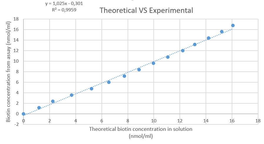
Figure 7: Plot of the theoretical amount of biotin in solution, versus the amount that's calculated based on the HABA-streptavidin assay.

Figure 8: Picture of end-results of the HABA-streptavidin test with, from left to right, a positive control (solution with excess biotin), the final cuvet of the concentration range of the PLA-biotin made with method 2, the final cuvet from method 3, the final cuvet of the concentration range of the untreated PLA and a negative control (nothing added).
After calculating the amounts of available biotin, the results are compared to each other in figure 9. This shows that the two filaments that were activated show a similar amount of biologically available biotin. The untreated PLA almost doesn’t show any change in absorbance.

Figure 9: measured amount of biologically available biotin per gram PLA for (from left to right); method 3, method 2 and untreated PLA
References
- Ben‐Shabat, S., Kumar, N., & Domb, A. J. (2006). PEG‐PLA Block Copolymer as Potential Drug Carrier: Preparation and Characterization. Macromolecular bioscience, 6(12), 1019-1025.
- Li, D., Frey, M. W., Vynias, D., & Baeumner, A. J. (2007). Availability of biotin incorporated in electrospun PLA fibers for streptavidin binding. Polymer, 48(21), 6340-6347.
- Salem, A. K., Cannizzaro, S. M., Davies, M. C., Tendler, S. J. B., Roberts, C. J., Williams, P. M., & Shakesheff, K. M. (2001). Synthesis and characterisation of a degradable poly (lactic acid)-poly (ethylene glycol) copolymer with biotinylated end groups. Biomacromolecules, 2(2), 575-580.
- Weiss, B., Schneider, M., Muys, L., Taetz, S., Neumann, D., Schaefer, U. F., & Lehr, C. M. (2007). Coupling of biotin-(poly (ethylene glycol)) amine to poly (D, L-lactide-co-glycolide) nanoparticles for versatile surface modification. Bioconjugate chemistry, 18(4), 1087-1094.