| Line 196: | Line 196: | ||
<a href="https://2016.igem.org/Team:BostonU_HW/Demonstrate">Demonstration</a> | <a href="https://2016.igem.org/Team:BostonU_HW/Demonstrate">Demonstration</a> | ||
<a href="https://2016.igem.org/Team:BostonU_HW/Proof">Proof</a> | <a href="https://2016.igem.org/Team:BostonU_HW/Proof">Proof</a> | ||
| − | <a href="https://2016.igem.org/Team:BostonU_HW/Design"> | + | <a href="https://2016.igem.org/Team:BostonU_HW/Design">Design</a> |
<a href="https://2016.igem.org/Team:BostonU_HW/Parts">Parts</a> | <a href="https://2016.igem.org/Team:BostonU_HW/Parts">Parts</a> | ||
</div> | </div> | ||
Revision as of 23:04, 19 October 2016
PROOF OF CONCEPT
Neptune (feat. HEK 293)
Putting Biology in Our Chips: Part 1
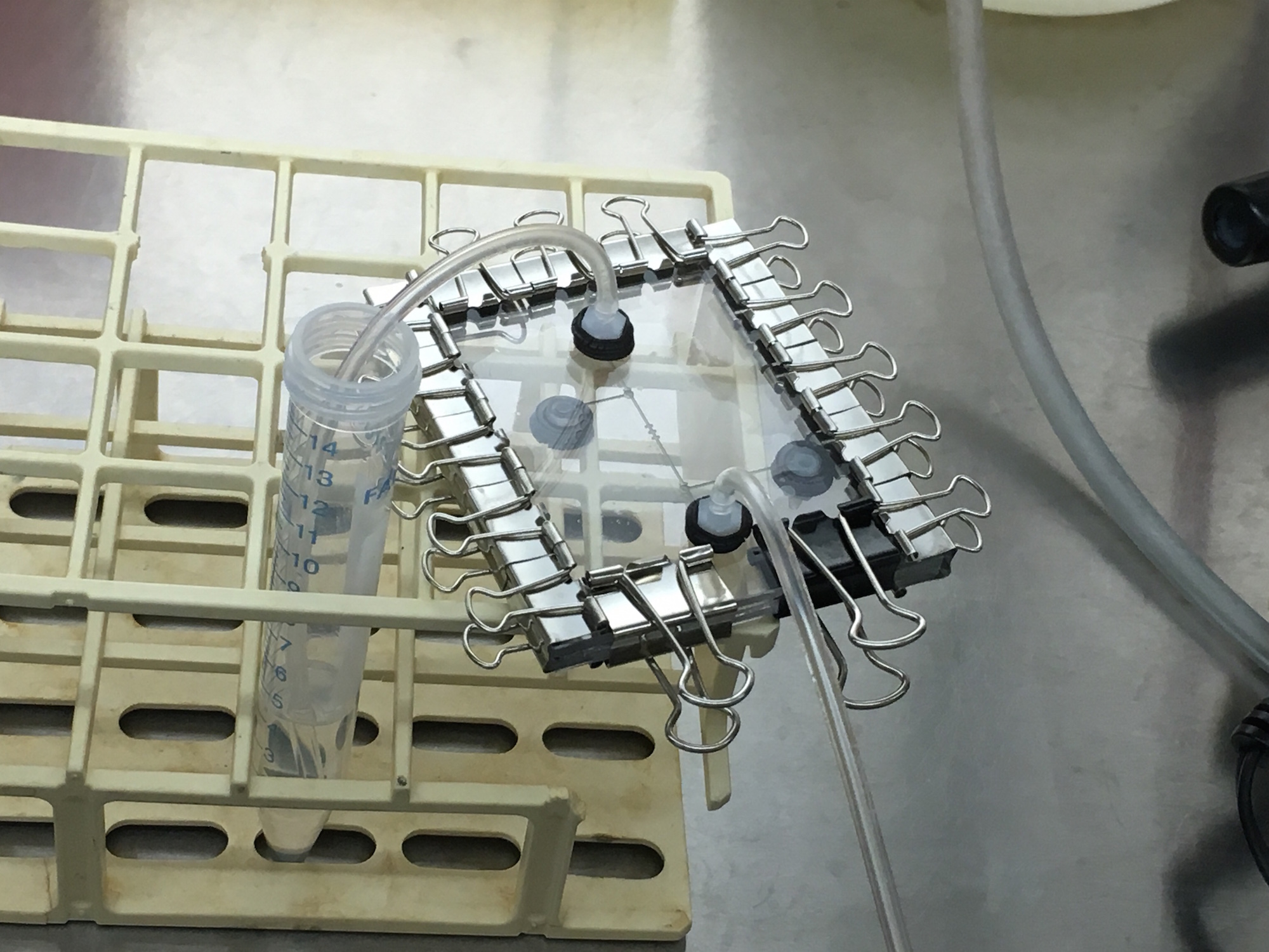
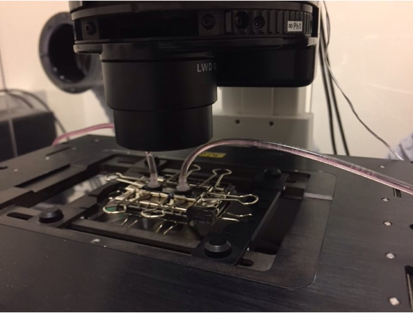
On our first visit to the MIT iGEM team's lab, we were testing a device with an input, and output, and a pair of valves surrounding cell traps in between. The design and fabrication of this device utilized our Neptune user interface, and proves it is capable of designing and manufacturing microfluidic devices for lab use. In the experiment (as further detailed on the collaborations page), we pulled ethanol solution, buffer solution, and cells through our microfluidic chip. In order to flow these liquids, we opened the (by default closed) valves with our servo-syringe combination pumps, utilizing our hardware and firmware in the process. The successful valve operation proves the proper functioning of our fluid controlling hardware, and the firmware which controls. After flowing cells through, we detached the microfluidic device from the control hardware (shutting the valves), and were able to see masses of HEK293 cells moving inside the valved off segment of the chip. Unfortunately, this evidence was only caught by eye and not on camera. However, we did capture images of the device under the microscope seen below.


Putting Biology in Our Chips: Part 2
Our second visit with the MIT iGEM team occurred within a week of the first and iterated on the previous design, highlighting the rapid prototyping made possible with the Neptune microfluidic workflow. This prototyping could have occurred even faster, as Neptune's the design and fabrication procedures take less than half a workday to complete, but both team's busy iGEM schedules were not that flexible. This design removed the controlling valves and pumps from the previous design, and replaced the cell traps with a diamond chamber meant to slow fluid flow at it's relatively wide center, allowing the HEK293 to attach to our device. Seen below, we captured evidence of the cells surviving and starting to multiple inside of our device. This is a proof of concept of mammalian cells (specifically HEK293) surviving and having the capacity to multiply within our devices.


Fictitious Synthetic Biologist: Dr. Ali
Our final example of our facilitated microfluidic workflow in action follows a fictitious researcher in synthetic biology, Dr. Ali. Dr. Ali is looking to automate and parallelize his ability to characterize a novel genetic part in terms of how cells express his reporter gene when various levels of inducer are present. The video below follows Dr. Ali through the entire process of creating a microfluidic device using the Neptune workflow, as well as controlling the chip with Neptune's parametric control hardware. This video gives a proof of concept for the entire user-interface driven design flow and also demonstrates how our control interface works in real time inconjunction with our servo-syringe control infrastructure.
{Disclaimer: The proof of concept microfluidic device seen in this video is demonstrating the ability to perform biological experiments, but actual biological components are not used. Instead, colored water is used as a model for the various components.}
{Disclaimer: The proof of concept microfluidic device seen in this video is demonstrating the ability to perform biological experiments, but actual biological components are not used. Instead, colored water is used as a model for the various components.}

