| Line 130: | Line 130: | ||
<u><p class="title1" id="select1">Predation</p></u> | <u><p class="title1" id="select1">Predation</p></u> | ||
| − | <p class=" | + | <p class="texte"> |
Our aim is to reinforce the natural predation capacity of <i>B. subtilis</i> and to ensure it is expressed independantly of the conditions. We | Our aim is to reinforce the natural predation capacity of <i>B. subtilis</i> and to ensure it is expressed independantly of the conditions. We | ||
| Line 158: | Line 158: | ||
<center><img src="https://static.igem.org/mediawiki/2016/2/28/Toulouse_France_results2.png" style="width:70%; margin:20px 20px;"> | <center><img src="https://static.igem.org/mediawiki/2016/2/28/Toulouse_France_results2.png" style="width:70%; margin:20px 20px;"> | ||
<b style="font-size:12px;"> | <b style="font-size:12px;"> | ||
| − | Figure 2 : Layout of SKF expected biobrick. | + | <br>Figure 2 : Layout of SKF expected biobrick. |
</b></center> | </b></center> | ||
<br><br> | <br><br> | ||
| Line 169: | Line 169: | ||
<center><img src="https://static.igem.org/mediawiki/2016/e/e0/Toulouse_France_results3.png" style="width:70%; margin:20px 20px;"> | <center><img src="https://static.igem.org/mediawiki/2016/e/e0/Toulouse_France_results3.png" style="width:70%; margin:20px 20px;"> | ||
<b style="font-size:12px;"> | <b style="font-size:12px;"> | ||
| − | Figure 3 : Layout of SDP expected biobrick. | + | <br>Figure 3 : Layout of SDP expected biobrick. |
</b></center><br><br> | </b></center><br><br> | ||
| Line 177: | Line 177: | ||
<center><img src="https://static.igem.org/mediawiki/2016/9/96/Toulouse_France_results4.png" style="width:15%; margin:20px 20px;"> | <center><img src="https://static.igem.org/mediawiki/2016/9/96/Toulouse_France_results4.png" style="width:15%; margin:20px 20px;"> | ||
<b style="font-size:12px;"> | <b style="font-size:12px;"> | ||
| − | Figure 4: Gibson assemby of the two SDP Gblocks. | + | <br>Figure 4: Gibson assemby of the two SDP Gblocks. |
</b></center><br><br> | </b></center><br><br> | ||
| Line 210: | Line 210: | ||
<center><img src="https://static.igem.org/mediawiki/2016/d/dc/Toulouse_France_results5.png" style="width:70%; margin:20px 20px;"> | <center><img src="https://static.igem.org/mediawiki/2016/d/dc/Toulouse_France_results5.png" style="width:70%; margin:20px 20px;"> | ||
<b style="font-size:12px;"> | <b style="font-size:12px;"> | ||
| − | Figure 5: Layout of antifungal operons and their assembly. | + | <br>Figure 5: Layout of antifungal operons and their assembly. |
</b></center><br><br> | </b></center><br><br> | ||
In order to express specifically the antifungal peptides in close vicinity to fungi, we choose the two N-acetyl-glucosamine (NAG) inducible promotors pNagA and pNagP. The constructions with the RFP reporter gene were ordered from IDT and successfully sub-cloned in the pSB1C3 (new parts <a href="http://parts.igem.org/Part:BBa_K1937003">BBa_K1937003</a> and <a href="http://parts.igem.org/Part:BBa_K1937005">BBa_K1937005</a> ; figure 6). They were subsequently cloned in the pSB<sub>BS</sub>0K-Mini plasmid to create biobricks <a href="http://parts.igem.org/Part:BBa_K1937004">BBA_K1937004</a> and <a href="http://parts.igem.org/Part:BBa_K1937006">BBa_K1937006</a>. | In order to express specifically the antifungal peptides in close vicinity to fungi, we choose the two N-acetyl-glucosamine (NAG) inducible promotors pNagA and pNagP. The constructions with the RFP reporter gene were ordered from IDT and successfully sub-cloned in the pSB1C3 (new parts <a href="http://parts.igem.org/Part:BBa_K1937003">BBa_K1937003</a> and <a href="http://parts.igem.org/Part:BBa_K1937005">BBa_K1937005</a> ; figure 6). They were subsequently cloned in the pSB<sub>BS</sub>0K-Mini plasmid to create biobricks <a href="http://parts.igem.org/Part:BBa_K1937004">BBA_K1937004</a> and <a href="http://parts.igem.org/Part:BBa_K1937006">BBa_K1937006</a>. | ||
| Line 218: | Line 218: | ||
<center><img src="https://static.igem.org/mediawiki/2016/4/41/Toulouse_France_results6.png" style="width:50%; margin:20px 20px;"> | <center><img src="https://static.igem.org/mediawiki/2016/4/41/Toulouse_France_results6.png" style="width:50%; margin:20px 20px;"> | ||
<b style="font-size:12px;"> | <b style="font-size:12px;"> | ||
| − | Figure 6: Layout of the pNag-RFP constructions. | + | <br>Figure 6: Layout of the pNag-RFP constructions. |
</b></center><br><br> | </b></center><br><br> | ||
| Line 230: | Line 230: | ||
<center><img src="https://static.igem.org/mediawiki/2016/4/4b/Toulouse_France_results7.png" style="width:40%; margin:20px 20px;"> | <center><img src="https://static.igem.org/mediawiki/2016/4/4b/Toulouse_France_results7.png" style="width:40%; margin:20px 20px;"> | ||
<b style="font-size:12px;"> | <b style="font-size:12px;"> | ||
| − | Figure 7: NAG-driven expression of RFP. <i>B. subtilis</i> strains transformed with pSB<sub>BS</sub>0K-Mini (Control), pSB<sub>BS</sub>0K-Mini-NagA or pSB<sub>BS</sub>0K-Mini-NagP were spread on minimal medium with either glucose or NAG as carbon source. Red spots appeared only with pNagA or pNagP on NAG (close-ups on part 7B). | + | <br>Figure 7: NAG-driven expression of RFP. <i>B. subtilis</i> strains transformed with pSB<sub>BS</sub>0K-Mini (Control), pSB<sub>BS</sub>0K-Mini-NagA or pSB<sub>BS</sub>0K-Mini-NagP were spread on minimal medium with either glucose or NAG as carbon source. Red spots appeared only with pNagA or pNagP on NAG (close-ups on part 7B). |
</b></center><br><br> | </b></center><br><br> | ||
| Line 244: | Line 244: | ||
<center><img src="https://static.igem.org/mediawiki/2016/9/99/Toulouse_France_results8.png" style="width:50%; margin:20px 20px;"> | <center><img src="https://static.igem.org/mediawiki/2016/9/99/Toulouse_France_results8.png" style="width:50%; margin:20px 20px;"> | ||
<b style="font-size:12px;"> | <b style="font-size:12px;"> | ||
| − | Figure 8: Antifungal tests (legend in the text). | + | <br>Figure 8: Antifungal tests (legend in the text). |
</b></center><br><br> | </b></center><br><br> | ||
| Line 275: | Line 275: | ||
<center><img src="https://static.igem.org/mediawiki/2016/4/49/Toulouse_France_results9.png" style="width:50%; margin:20px 20px;"> | <center><img src="https://static.igem.org/mediawiki/2016/4/49/Toulouse_France_results9.png" style="width:50%; margin:20px 20px;"> | ||
<b style="font-size:12px;"> | <b style="font-size:12px;"> | ||
| − | Figure 9: Layout of the toxin/antitoxin operons. | + | <br>Figure 9: Layout of the toxin/antitoxin operons. |
</b></center><br><br> | </b></center><br><br> | ||
| Line 286: | Line 286: | ||
<center><img src="https://static.igem.org/mediawiki/2016/5/5b/Toulouse_France_results10.png" style="width:40%; margin:20px 20px;"> | <center><img src="https://static.igem.org/mediawiki/2016/5/5b/Toulouse_France_results10.png" style="width:40%; margin:20px 20px;"> | ||
<b style="font-size:12px;"> | <b style="font-size:12px;"> | ||
| − | + | <br>sFigure 10: Result of the <i>Bacillus subtilis</i> transformation with pSB<sub>BS</sub>0K-Mini –Epsilon/MazF (we know this is not the most illustrative figure ever !). | |
</b></center><br><br> | </b></center><br><br> | ||
Revision as of 20:30, 19 October 2016
Results
Predation
Our aim is to reinforce the natural predation capacity of B. subtilis and to ensure it is expressed independantly of the conditions. We
first assessed that our wild type Bacillus chassis is not able of predation, then we built the operons
allowing boosting the predation property.
Preliminary tests:
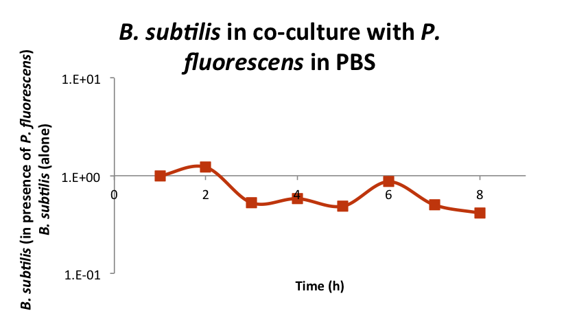
We tried different testing approaches to evaluate the predatory response of B. subtilis and eventually elaborate a protocol to do the preliminary tests. We tested the predation of B. subtilis Wild Type (strain 168) against Pseudomonas fluorescens (strain SBW25), a deleterious strain present in the cave. Briefly, the protocol consists in growing both strains in rich medium, mixing them in PBS and monitoring their growth (figure 1).

Figure 1: Bacillus subtilis WT 168 does not feed on Pseudomonas fluorescens SBW25. Both strains were grown overnight in rich medium and then mixed in PBS. The growths of the strains were then monitored during 8 hours by plate numeration. The graph represents the ratio between B. subtilis in PBS in presence of P. fluorescens versus B. subtilis alone in PBS (data normalized to time 1H).
We observed no growth benefit when mixing B. subtilis and P. fluorescens compared to B. subtilis alone. We conclude that B. subtilis WT predation program is not the strain priority when facing starvation. Other surviving program as competence or sporulation are likely favoured by B. subtilis in such condition. This reinforces the need to prevent these programs by using a spo0A mutant and to promote the predation by overexpressing either the SKF or SDP operons.
SKF
This predation operon is composed of seven genes for a total of more than 6 kb. To get rid of restriction sites that could interfere with the cloning steps, we ordered the optimized sequences from IDT as four gblocks. From there, our strategy was to do Gibson cloning to obtain the full operon in pSB1C3 in E. coli and then to transfer it in B. subtilis. However, we did not manage to obtain the whole assembly (figure 2), neither partial ones, in spite of about 20 attempts…

Figure 2 : Layout of SKF expected biobrick.
SDP
The SDP operon is smaller than the SKF one and it was possible to obtain the optimized sequences as two gblocks. Here again, we were unfortunate and did not get the expected clones in E. coli (figure 3).

Figure 3 : Layout of SDP expected biobrick.
To perform trouble shooting, we tried an assembly test with just the two gblocks and deposited the product on gel. We observed that the reaction seems to be effective with the presence of a new band corresponding to the combined size of the two gblocks (figure 4).

Figure 4: Gibson assemby of the two SDP Gblocks.
Conclusions and perspectives
It seems our Gibson step is fine since we managed to obtain the SDP assembly, but we could not get E. coli transformants when performing the whole experience. The predation system is based on the production of toxins by B. subtilis, and these toxins were reported to be harmful to E. coli (Nandy et al., 2007, FEBS Letters. 581: 151–56). An explanation to our problems could be that SDP and SKF cloning in E. coli results in the bacterium death. We had thought about this problem, but we had believed the expression driven by the pVeg Bacillus promoter to be insufficient for such effect. Perspectives could be to use a tightly regulated promoter to prevent expression during the cloning step in E. coli, or to try a direct transformation of highly competent Bacillus strain.
Antifungals
Here, we aimed to produce a cocktail of five antifungal peptides whose production in Bacillus subtilis will be triggered by presence of fungi.
Operon constructions:
The whole antifungal operon was too big to be synthesized by IDT as one gblock. We therefore decided to divide it in two operons (figure 5), each of them with a promoter to be functional, with the possibility to eventually combine them. The sequence were optimized for the Bacillus codon usage and to remove inadequate restriction sites. Sub-cloning of the first operon (containing cut version of the Metchnikowin and D4E1) on the pSB1C3 backbone was rapidly performed, leading to the new composite part BBa_K1937007 (pSB1C3-AF_A). However, we did not manage to obtain the second operon in the pSB1C3 (encoding Dermaseptin B1, GAFP-1 and entire Metchnikowin antifungal peptides). We tried to directly sub-clone the gblock in the pSB1C3-AF_A but without success. We hypothesize that one of the peptide could be toxic for E. coli. This will have to be verified by sub-cloning the 3 peptides alone. The AF_A operon was subsequently cloned in the pSBBS0K-Mini plasmid to create biobrick BBA_K1937008.

Figure 5: Layout of antifungal operons and their assembly.
In order to express specifically the antifungal peptides in close vicinity to fungi, we choose the two N-acetyl-glucosamine (NAG) inducible promotors pNagA and pNagP. The constructions with the RFP reporter gene were ordered from IDT and successfully sub-cloned in the pSB1C3 (new parts BBa_K1937003 and BBa_K1937005 ; figure 6). They were subsequently cloned in the pSBBS0K-Mini plasmid to create biobricks BBA_K1937004 and BBa_K1937006.

Figure 6: Layout of the pNag-RFP constructions.
pNag validation
We tested the expression and specificity of the RFP driven by pNagA and pNagP when growing in presence of glucose or NAG (figure 7). We observed a late and rather specific RFP expression on NAG. The late expression could mean that the formulation of our minimal medium is not optimal. The fact that the pNagA-RFP and pNagP-RFP strains seem able to slightly express the RFP on glucose (figure 7B, left panel close-up), albeit on weaker extend that on NAG (figure 7B, right panel close-up), could be due to the alleviating of the catabolic repression.
In conclusion, pNAgA and pNagP appear as able to promote expression in response to NAG, even if the growth conditions could be improved to get higher and more homogeneous expression levels.

Figure 7: NAG-driven expression of RFP. B. subtilis strains transformed with pSBBS0K-Mini (Control), pSBBS0K-Mini-NagA or pSBBS0K-Mini-NagP were spread on minimal medium with either glucose or NAG as carbon source. Red spots appeared only with pNagA or pNagP on NAG (close-ups on part 7B).
Antifungal validation
We found out that the best culture conditions for the fungi that permits a slight growth of Bacillus were with ¼ PDA and 2% glucose. We tested different fungi (Aspergillus niger, Talaromyces funiculosus and Chaetomium globosum) but we eventually focussed on Talaromyces funiculosus that seems easier to manipulate to us.
Our test consisted in adding, on fungi inoculated plates, paper patches soaked with either copper sulfate (positive control), LB medium (negative control), a suspension of Bacillus subtilis WT or Bacillus subtilis expressing the antifungal AF_A operon (figure 8). We observed that with our construction, a slight inhibition halo appeared around the patch. This effect is visible even after 8 days and was reproducible. These observations allow us to conclude that AF_A is functional.

Figure 8: Antifungal tests (legend in the text).
Conclusions and perspectives
Here, we showed that our pNagA and NagP parts are able to control gene expression in response to NAG and that the first part of our antifungal operon is functional. In both cases, the properties will have to be optimized, through a higher and more homogeneous expression from the NAG-driven promoters and through the completion of the antifungal operons to produce more than two antifungal peptides.
Confinement
Here, we fashioned a genetic system to prevent horizontal transfer of our synthetic constructions.
Toxin/antitoxin systems constructions
The constructions were ordered as gblocks from IDT. The Epsilon/MazF construction was rapidly sub-cloned in the pSB1C3 backbone (new composite part BBa_K1937007), and then in the pSBBS0K-Mini plasmid to create biobricks BBA_K1937008 (figure 9). However, we never managed to get the MazE/Zeta construction in the pSB1C3 backbone. Again, we can only speculate about the toxicity of the toxin.

Figure 9: Layout of the toxin/antitoxin operons.
Theophylline validation
To validate the theophylline riboswitch, we inferred that we should obtain clones of Bacillus subtilis transformed with the pSBBS0K-Mini –Epsilon/MazF only in presence of theophylline: the molecule should prevent the expression of the MazF toxin that is lethal since the antitoxin MazE is not present. Unfortunately, we did not get any clone, neither without nor with theophylline (figure 10).

sFigure 10: Result of the Bacillus subtilis transformation with pSBBS0K-Mini –Epsilon/MazF (we know this is not the most illustrative figure ever !).
Conclusions and perspectives
At this step, we can only hypothesize that our system is leaking sufficient expression of the toxins for them to be lethal, either in E. coli or in B. subtilis. Further assays using inducible promoters will be necessary to set up the system without enduring these toxicity problems.

Website by Team iGEM Toulouse 2016


