| Line 34: | Line 34: | ||
<ul> | <ul> | ||
<li>When our constructs were in DH10B cells, the <i>fldA</i>, <i>fldA</i>-<i>pfo</i>, <i>petF</i>, and <i>petF</i>-<i>pfo</i> strains produced more biotin than the control DH10B strain at some induction level</li> | <li>When our constructs were in DH10B cells, the <i>fldA</i>, <i>fldA</i>-<i>pfo</i>, <i>petF</i>, and <i>petF</i>-<i>pfo</i> strains produced more biotin than the control DH10B strain at some induction level</li> | ||
| − | <li>When our constructs were in the Δ<i>aceE</i> knockout cells, the <i>fldA</i>, <i>fldA</i>-<i>pfo</i>, and <i>petF</i> strains all produced more biotin than the control DH10B strain, control Δ<i>aceE</i> knockout strain, and DH10B+constructs strain (<i>petF</i>-<i>pfo</i> data is | + | <li>When our constructs were in the Δ<i>aceE</i> knockout cells, the <i>fldA</i>, <i>fldA</i>-<i>pfo</i>, and <i>petF</i> strains all produced more biotin than the control DH10B strain, control Δ<i>aceE</i> knockout strain, and DH10B+constructs strain (<i>petF</i>-<i>pfo</i> data is not present because it was not able to grow)</li> |
</ul> | </ul> | ||
</p> | </p> | ||
| Line 41: | Line 41: | ||
<img src = "https://static.igem.org/mediawiki/2016/c/c5/T--WashU_StLouis--BiotinYalegraph.png" style="width : 40vw;display: inline-block; "> | <img src = "https://static.igem.org/mediawiki/2016/c/c5/T--WashU_StLouis--BiotinYalegraph.png" style="width : 40vw;display: inline-block; "> | ||
</div> | </div> | ||
| + | |||
| + | <p>Additionally, we saw a phenotypic change in our electron donor cells. Post-induction, the cells that contained the <i>petF</i> or <i>petF</i>-<i>pfo</i> constructs had a distinct red color. The red color is likely due to the increased amounts of ferredoxin in the cell, which <i>E. coli</i> is obviously not use to. The color may come from a large amount of cytochrome d, another electron transport protein which has red color when it is reduced[3].</p> | ||
| + | |||
| + | ADD RED CELL PHOTOS HERE! THERE SHOULD BE 2 - ONE FOR DH10B AND ONE FOR YALE CELLS | ||
<p>This experiment illustrates a proof of concept for our Super Cells. By showing that our cells could overproduce biotin, we have shown that the theory behind the Super Cells work: that overproducing certain co-factors will increase the amount of resulting product<p> | <p>This experiment illustrates a proof of concept for our Super Cells. By showing that our cells could overproduce biotin, we have shown that the theory behind the Super Cells work: that overproducing certain co-factors will increase the amount of resulting product<p> | ||
| Line 57: | Line 61: | ||
<li>Lin, Steven, and John E. Cronan. "Closing in on complete pathways of biotin biosynthesis." Molecular BioSystems 7.6 (2011): 1811-1821. </li> | <li>Lin, Steven, and John E. Cronan. "Closing in on complete pathways of biotin biosynthesis." Molecular BioSystems 7.6 (2011): 1811-1821. </li> | ||
<li>Picciocchi, Antoine, Roland Douce, and Claude Alban. "Biochemical characterization of the Arabidopsis biotin synthase reaction. The importance of mitochondria in biotin synthesis." Plant physiology 127.3 (2001): 1224-1233.</li> | <li>Picciocchi, Antoine, Roland Douce, and Claude Alban. "Biochemical characterization of the Arabidopsis biotin synthase reaction. The importance of mitochondria in biotin synthesis." Plant physiology 127.3 (2001): 1224-1233.</li> | ||
| + | <li>Haddock, B. A., J. ALLAN Downie, and P. B. Garland. "Kinetic characterization of the membrane-bound cytochromes of Escherichia coli grown under a variety of conditions by using a stopped-flow dual-wavelength spectrophotometer." Biochemical Journal 154.2 (1976): 285-294.</li> | ||
</ol> | </ol> | ||
</div> | </div> | ||
Revision as of 22:32, 18 October 2016
Proof of Concept
Measuring biotin production to calculate reduced electron donors

After analyzing the results of our absorbance data of electron donors, we decided to go a step further to ensure that our electron donor constructs were producing reduced and usable electron donors. We performed an assay which showed that not only were the products being overproduced, but also that there was significant data supporting the claim that these electron donors were usable in protein synthesis.
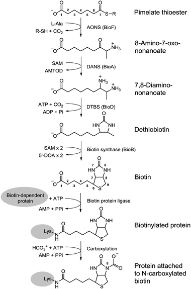
Biotin, commonly know as vitamin B7, is produced by a pathway that uses reduced flavodoxin/ferredoxin,specifically the SAM pathway that turns dethiobiotin to biotin[1] (see diagram below). Specifically, there is a 1:1 stoichiometric relationship between reduced electron donors and biotin molecules produced. If we measure the amount of biotin in our cells, we will indirectly measure the amount of reduced electron donors present in our cells. We used a readily available biotin assay to conduct our proof of concept experiment.
Results showed that overall, our constructs worked to increase biotin production, as seen in the graphs below.
- When our constructs were in DH10B cells, the fldA, fldA-pfo, petF, and petF-pfo strains produced more biotin than the control DH10B strain at some induction level
- When our constructs were in the ΔaceE knockout cells, the fldA, fldA-pfo, and petF strains all produced more biotin than the control DH10B strain, control ΔaceE knockout strain, and DH10B+constructs strain (petF-pfo data is not present because it was not able to grow)


Additionally, we saw a phenotypic change in our electron donor cells. Post-induction, the cells that contained the petF or petF-pfo constructs had a distinct red color. The red color is likely due to the increased amounts of ferredoxin in the cell, which E. coli is obviously not use to. The color may come from a large amount of cytochrome d, another electron transport protein which has red color when it is reduced[3].
ADD RED CELL PHOTOS HERE! THERE SHOULD BE 2 - ONE FOR DH10B AND ONE FOR YALE CELLSThis experiment illustrates a proof of concept for our Super Cells. By showing that our cells could overproduce biotin, we have shown that the theory behind the Super Cells work: that overproducing certain co-factors will increase the amount of resulting product
Go to the conclusions page to see a complete summary of our results and proof of concept experiments.
References
- Lin, Steven, and John E. Cronan. "Closing in on complete pathways of biotin biosynthesis." Molecular BioSystems 7.6 (2011): 1811-1821.
- Picciocchi, Antoine, Roland Douce, and Claude Alban. "Biochemical characterization of the Arabidopsis biotin synthase reaction. The importance of mitochondria in biotin synthesis." Plant physiology 127.3 (2001): 1224-1233.
- Haddock, B. A., J. ALLAN Downie, and P. B. Garland. "Kinetic characterization of the membrane-bound cytochromes of Escherichia coli grown under a variety of conditions by using a stopped-flow dual-wavelength spectrophotometer." Biochemical Journal 154.2 (1976): 285-294.


