TrinhNguyen (Talk | contribs) |
TrinhNguyen (Talk | contribs) |
||
| Line 28: | Line 28: | ||
<h2 style="color: #F20253; text-decoration:underline; font-family: Trebuchet MS;">Purpose</h2> | <h2 style="color: #F20253; text-decoration:underline; font-family: Trebuchet MS;">Purpose</h2> | ||
| − | <a style="text-decoration: none; color: #000000; float: | + | <a style="text-decoration: none; color: #000000; float: left; margin: 15px"> |
| − | <img src="https://static.igem.org/mediawiki/2016/c/c4/T--MIT--microfluidic_device.jpeg" alt="Microfluidic device"/ style="width: | + | <img src="https://static.igem.org/mediawiki/2016/c/c4/T--MIT--microfluidic_device.jpeg" alt="Microfluidic device"/ style="width:200px; float: left" margin: 15px;> |
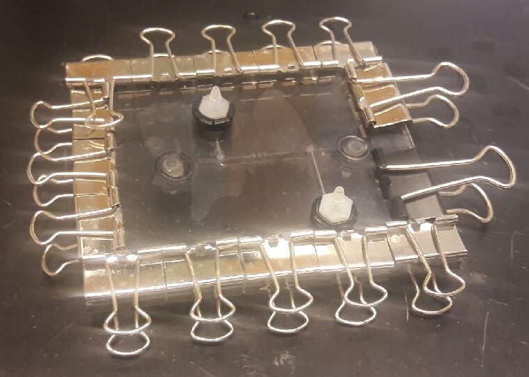
<div style='width: 340px; text-align: center;'><i>Microfluidic device fabricated by BU Hardware iGEM Team.</i></div> | <div style='width: 340px; text-align: center;'><i>Microfluidic device fabricated by BU Hardware iGEM Team.</i></div> | ||
</a> | </a> | ||
| Line 38: | Line 38: | ||
</p> | </p> | ||
| − | <p style="font-weight:bold; font-family: | + | <p style="font-weight:bold; font-family:Verdana;">How does microfluidics benefit our project?</p> |
<ol style="font-family:Verdana;"> | <ol style="font-family:Verdana;"> | ||
<li>Automatically regulate estrogen and progestorone cycle in vitro cell culture</li> | <li>Automatically regulate estrogen and progestorone cycle in vitro cell culture</li> | ||
Revision as of 03:07, 18 October 2016
Collaborations
Mammalian cell culture in microfluidic devices
Purpose

Integration of microfluidic devices has advanced considerably in quantitative biological research [1][2]. The timing of the fluid flow can be regulated with high accuracy because of the short response time of in-chip membrane valves (as quick as 1ms)[1]. Moreover, with the aid of hydrogel, cells growing in microfluidics can be structured in three-dimension geometries [1]. Therefore, cell culture performed in microfluidic devices could resemble in vivo conditions due to tight control of microenvironment [2]. On the other hand, automation is another advantageous of microfluidic technology. Instead of manual intervention, programmable fluid flows can automatically change chemical conditions of the culture environment. This feature would be particularly useful to examine dynamically changing systems [1]. The ability to precisely control fluid flows and chemical species, and automation of microfluidic technology are highly beneficial to future development of our project. Thus, MIT iGEM team decided to collaborate with the Boston University Hardware iGEM team to develop a protocol for mammalian cell culture in microfluidic devices
How does microfluidics benefit our project?
- Automatically regulate estrogen and progestorone cycle in vitro cell culture
- Resemble dynamically changing estrogen and progesterone concentration
- Dynamically monitor cell reponse and output gene expression
Collaboration
We collaborated with the Boston University Hardware iGEM Team to culture mammalian cells in their microfluidic devices. We started with the simplest experiment, which is culturing HEK293 (Human Embryonic Kidney) cells in a microfluidic device. We run the test experiment with two different models of the microfluidic device.
Model 1
Microfluidic device
The device had 2 sets of inlet-outlet channels, one for cells seeding, and the other set for controling chemical species' concentrations in media. The first model did not have any chamber or a specific region for cell cultureing. The inlet and outlet were connected by a tube with uniform dimensions. It, however, had rectangular cell traps along the channel.
Cell culturing protocol
Model 2
Microfluidic device
Since we couldn't find any cells under 10X microscope in the first-model device, we proposed several modifications for the device to the BU hardware team after doing some literature research, which included:
- Create a diamond-shape chamber, a specific region for cells adhesion. The increase in cross-section area along the channel reduces the flow rate of the fluid, making it easier the cells to stick to the surface of the chamber.
- Using a syringe pump to continuously replenish the media inside the chamber after cell adhesion, preventing the media from drying out inside the chamber - which happened in model 1.
Cell culturing protocol
Results and discussion
Testing pDEST mCherry
Purpose
Collaboration
Results
Recombinases' sequences
Purpose
Collaboration
Results
REFERENCE:
- Mehling Matthias, and Tay Savas. Microfluidic Cell Culture. Science Direct 2014; 25:95-102.
- Komen Job, Wolbers Floor, Franke R. Henk, et al. Viability analysis and apoptosis induction of breast cancer cells in a microfluidic device: effect of cytostatic drugs. Biomed Microdevices(2008); 10:727–737


