(→Hypoxia-inducible promoters) |
(→Fighting hypoxia in printed tissue) |
||
| (43 intermediate revisions by 5 users not shown) | |||
| Line 1: | Line 1: | ||
| − | {{LMU-TUM_Munich|navClass= | + | {{LMU-TUM_Munich|navClass=hypoxia}} |
| − | + | ||
__NOTOC__ | __NOTOC__ | ||
[[File:Muc16_Sticker_Hypoxia_001.png |right|350px]] | [[File:Muc16_Sticker_Hypoxia_001.png |right|350px]] | ||
| Line 8: | Line 7: | ||
<div class="white-box"> | <div class="white-box"> | ||
| − | Unicellular organisms such as bacteria | + | Unicellular organisms such as bacteria possess the ability to reproduce very quickly when given the right conditions such as warmth, moisture and suitable nutrients,<ref>http://www.bbc.co.uk/schools/gcsebitesize/science/ |
| + | triple_ocr_gateway/beyond_the_microscope/understanding_microbes/revision/2/</ref> and enter an inactive state when these are limited. On the contrary, most multicellular systems depend on a steady supply of oxygen and nutrients essential for their survival and metabolic stability. | ||
| − | Herein lies one of the main challenges of bioprinting approaches. | + | Herein lies one of the main challenges of [https://2016.igem.org/Team:LMU-TUM_Munich/Demonstrate bioprinting approaches]. |
When printing, the homogenous delivery of oxygen to three-dimensional cell-constructs is limited by diffusion gradients in tissue from the periphery towards the center <ref>Volkmer, Elias, et al. "Hypoxia in static and dynamic 3D culture systems for tissue engineering of bone." Tissue Engineering Part A 14.8 (2008): 1331-1340</ref>, thus resulting in tissue hypoxia. <ref> Höckel, Michael, and Peter Vaupel. "Tumor hypoxia: definitions and current clinical, biologic, and molecular aspects." Journal of the National Cancer Institute 93.4 (2001): 266-276. </ref> | When printing, the homogenous delivery of oxygen to three-dimensional cell-constructs is limited by diffusion gradients in tissue from the periphery towards the center <ref>Volkmer, Elias, et al. "Hypoxia in static and dynamic 3D culture systems for tissue engineering of bone." Tissue Engineering Part A 14.8 (2008): 1331-1340</ref>, thus resulting in tissue hypoxia. <ref> Höckel, Michael, and Peter Vaupel. "Tumor hypoxia: definitions and current clinical, biologic, and molecular aspects." Journal of the National Cancer Institute 93.4 (2001): 266-276. </ref> | ||
| Line 18: | Line 18: | ||
Sprouting angiogenesis, as implied by its name is characterized by sprouts composed of endothelial cells, which usually grow toward an angiogenic stimulus. | Sprouting angiogenesis, as implied by its name is characterized by sprouts composed of endothelial cells, which usually grow toward an angiogenic stimulus. | ||
| − | [[File:VEGF.png|thumb|right|450px| | + | [[File:VEGF.png|thumb|right|450px| <b>Figure 1:</b> Schematic depiction of the VEGF receptor with bound growth factor, triggering vascularization.<ref>Modified from: http://www.liquidarea.com/2011/05/al-cnr-arrivano-i-farmaci-intelligenti-terapie-del-futuro/</ref>]] |
| − | + | ||
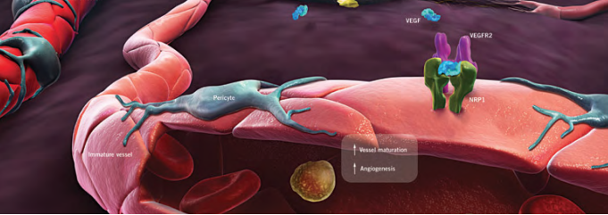
This driving stimulus is initiated in poorly perfused tissues when oxygen sensing mechanisms detect a level of hypoxia that demands the formation of new blood vessels to satisfy the metabolic requirements of parenchymal cells. Most types of parenchymal cells respond to a hypoxic environment by secreting a key proangiogenic growth factor called vascular endothelial growth factor (VEGF-A). <ref>Adair TH, Montani JP. Angiogenesis. San Rafael (CA): Morgan & Claypool Life Sciences; 2010. Chapter 1, Overview of Angiogenesis | This driving stimulus is initiated in poorly perfused tissues when oxygen sensing mechanisms detect a level of hypoxia that demands the formation of new blood vessels to satisfy the metabolic requirements of parenchymal cells. Most types of parenchymal cells respond to a hypoxic environment by secreting a key proangiogenic growth factor called vascular endothelial growth factor (VEGF-A). <ref>Adair TH, Montani JP. Angiogenesis. San Rafael (CA): Morgan & Claypool Life Sciences; 2010. Chapter 1, Overview of Angiogenesis | ||
doi:10.1089/ten.tea.2007.0231</ref> | doi:10.1089/ten.tea.2007.0231</ref> | ||
| − | Because the production of a normal vasculature is among others heavily dependent upon the concentration of VEGF-A in the tissue, we have constructed a model in order to measure the hypoxia dependent secretion of VEGF and PDGF, | + | Because the production of a normal vasculature is among others heavily dependent upon the concentration of VEGF-A in the tissue, we have constructed a model in order to measure the hypoxia dependent secretion of VEGF and PDGF (the so-called ''platelet-derived growth factor'', which stimualtes cell division<ref>Li, X., & Eriksson, U. (2003). Novel PDGF family members: PDGF-C and PDGF-D. Cytokine & growth factor reviews, 14(2), 91-98.</ref>). Thus, we may simulate the extreme conditions that are present when printing several layers of tissue and, by triggering sprouting angiogenesis, induce growth of blood vessels towards portions of tissues previously devoid of them. |
</div> | </div> | ||
| Line 32: | Line 31: | ||
Hypoxia refers to subnormal levels of oxygen in air, blood and tissue. Tissue hypoxia leads to cellular dysfunction and can ultimately lead to cell death. The ability of cells to adapt to periods of hypoxia is therefore important for their survival. <ref>Muz, Barbara, et al. "Hypoxia. The role of hypoxia and HIF-dependent signalling events in rheumatoid arthritis." Arthritis research & therapy 11.1 (2009): 1</ref> | Hypoxia refers to subnormal levels of oxygen in air, blood and tissue. Tissue hypoxia leads to cellular dysfunction and can ultimately lead to cell death. The ability of cells to adapt to periods of hypoxia is therefore important for their survival. <ref>Muz, Barbara, et al. "Hypoxia. The role of hypoxia and HIF-dependent signalling events in rheumatoid arthritis." Arthritis research & therapy 11.1 (2009): 1</ref> | ||
| − | In this context, we wanted to find the most suitable way to compensate the poor oxygen conditions that are generated when printing tissue structures with a volume of more than | + | In this context, we wanted to find the most suitable way to compensate the poor oxygen conditions that are generated when printing tissue structures with a volume of more than a few mm<sup>3</sup>. |
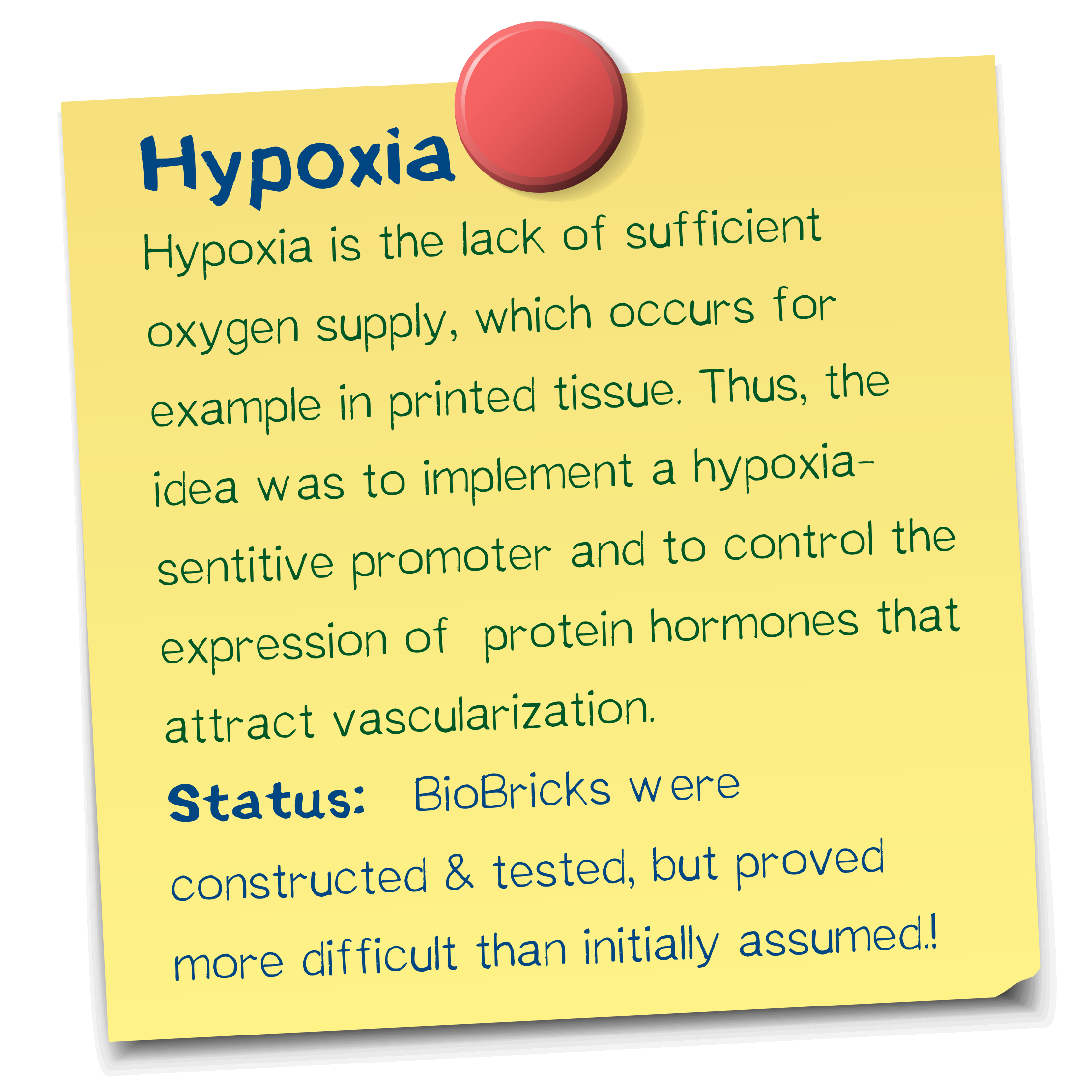
Following a previously published idea by Lee, J. Y., et al. "A novel chimeric promoter that is highly responsive to hypoxia and metals." to enhance the level of hypoxic induction and to control target gene expression <ref>Lee, J. Y., et al. "A novel chimeric promoter that is highly responsive to hypoxia and metals." Gene therapy 13.10 (2006): 857-868</ref>, we have designed a potent hypoxia-inducible promoter system, making use of so-called hypoxia-response elements. | Following a previously published idea by Lee, J. Y., et al. "A novel chimeric promoter that is highly responsive to hypoxia and metals." to enhance the level of hypoxic induction and to control target gene expression <ref>Lee, J. Y., et al. "A novel chimeric promoter that is highly responsive to hypoxia and metals." Gene therapy 13.10 (2006): 857-868</ref>, we have designed a potent hypoxia-inducible promoter system, making use of so-called hypoxia-response elements. | ||
Many hypoxia-responsive genes are regulated by the hypoxia-response element (HRE) protein family of transcription factors, including hypoxia-inducible factor-1 (HIF-1). <ref>Lee, J. Y., et al. "A novel chimeric promoter that is highly responsive to hypoxia and metals." Gene therapy 13.10 (2006): 857-868</ref> | Many hypoxia-responsive genes are regulated by the hypoxia-response element (HRE) protein family of transcription factors, including hypoxia-inducible factor-1 (HIF-1). <ref>Lee, J. Y., et al. "A novel chimeric promoter that is highly responsive to hypoxia and metals." Gene therapy 13.10 (2006): 857-868</ref> | ||
| − | When HREs derived from different genes are placed in plasmids, they confer hypoxia inducibility upon heterologous promoters in various cell types. <ref>Boast K, Binley K, Iqball S, Price T, Spearman H, Kingsman S et al. Characterization of physiologically regulated vectors for the treatment of ischemic disease. Hum Gene Ther 1999; 10: 2197–2208</ref> | + | When HREs derived from different genes are placed in plasmids, they confer hypoxia inducibility upon heterologous promoters in various cell types. <ref>Boast K, Binley K, Iqball S, Price T, Spearman H, Kingsman S et al. Characterization of physiologically regulated vectors for the treatment of ischemic disease. Hum Gene Ther 1999; 10: 2197–2208</ref> We intend to use this system of hypoxia-inducible expression to induce vascularization in printed tissues. |
</div> | </div> | ||
| Line 45: | Line 44: | ||
<div class="white-box"> | <div class="white-box"> | ||
| − | In order to test the responsivity of HREs we incorporated different copy numbers (1x-HRE, 6x-HRE, 8x-HRE) upstream from a minimal promoter. The evaluation of the constructs inductivity was performed by measuring the secretion of a luciferase reporter after transient transfection in HEK293T cells | + | In order to test the responsivity of HREs, we incorporated different copy numbers (1x-HRE, 6x-HRE, 8x-HRE) upstream from a minimal promoter. The evaluation of the constructs' inductivity was performed by measuring the secretion of a luciferase reporter after transient transfection in HEK293T cells. |
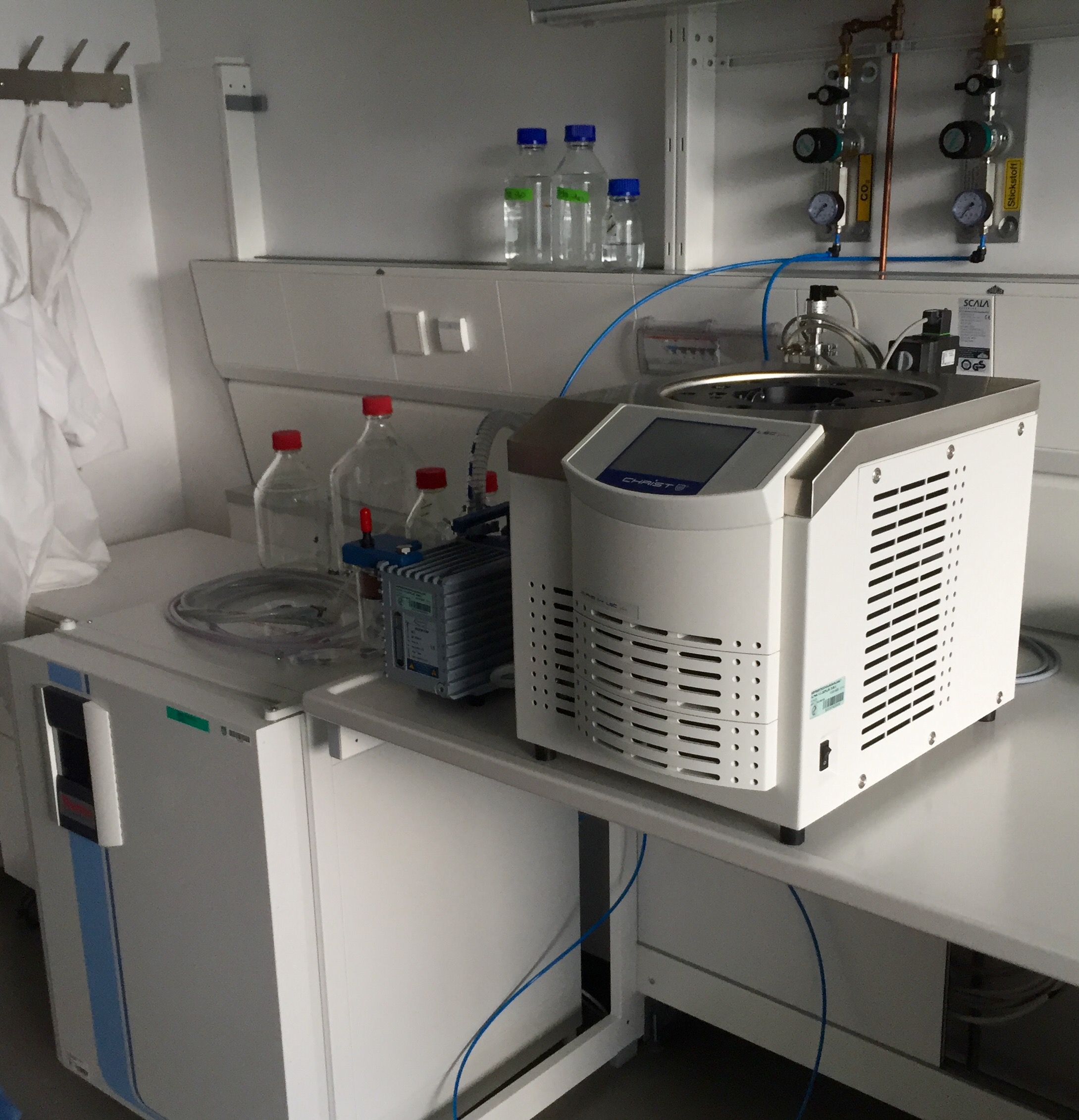
| − | Parallely we expressed a HRE´s activated construct containing the actual growth factors VEGF and PDGF under the same conditions | + | [[File:Inkubator2.jpeg|thumb|right|300px| <i><b>Figure 2:</b></i> The incubation facility at the ''Max-Planck-Institute for Biochemistry'' waiting to be tested.]] |
| − | + | ||
| + | Parallely we expressed a HRE´s activated construct containing the actual growth factors VEGF and PDGF under the same conditions and compared it to a positive control consisting of a CMV driven VEGF and PDGF gene. | ||
For the experiments cells were subjected to normoxia (21% O<sub>2</sub>, 5% CO<sub>2</sub>, 74% N<sub>2</sub>) and to hypoxia (1% O<sub>2</sub>, 7% CO<sub>2</sub>, 92% N<sub>2</sub>) for 24 hours. | For the experiments cells were subjected to normoxia (21% O<sub>2</sub>, 5% CO<sub>2</sub>, 74% N<sub>2</sub>) and to hypoxia (1% O<sub>2</sub>, 7% CO<sub>2</sub>, 92% N<sub>2</sub>) for 24 hours. | ||
| − | |||
| − | |||
By taking samples of the medium after 0 h, 12 h and 24 h after the exposure of the cells to this conditions and measuring luciferase activity by a corresponding assay, we were able to quantify hypoxia-dependent gene expression. To be able to determine how far gene expression is responsive to hypoxia in comparison to normal oxygen concentrations, a control at normoxic conditions was furthermore run. | By taking samples of the medium after 0 h, 12 h and 24 h after the exposure of the cells to this conditions and measuring luciferase activity by a corresponding assay, we were able to quantify hypoxia-dependent gene expression. To be able to determine how far gene expression is responsive to hypoxia in comparison to normal oxygen concentrations, a control at normoxic conditions was furthermore run. | ||
| − | For the assay, a total of five constructs were tested, three of which were luciferase constructs for the quantification of expression levels and two of which were VEGF-constructs that aimed to confirm the expression of the actual growth factor by our cells, as well as acting as a negative control for the luciferase assay. The luciferase constructs all contained the hypoxia promoter. | + | For the assay, a total of five constructs were tested, three of which were luciferase constructs for the quantification of expression levels and two of which were VEGF-constructs that aimed to confirm the expression of the actual growth factor by our cells via ELISA, as well as acting as a negative control for the luciferase assay. The luciferase constructs all contained the hypoxia promoter. |
| Line 64: | Line 62: | ||
<div class="white-box"> | <div class="white-box"> | ||
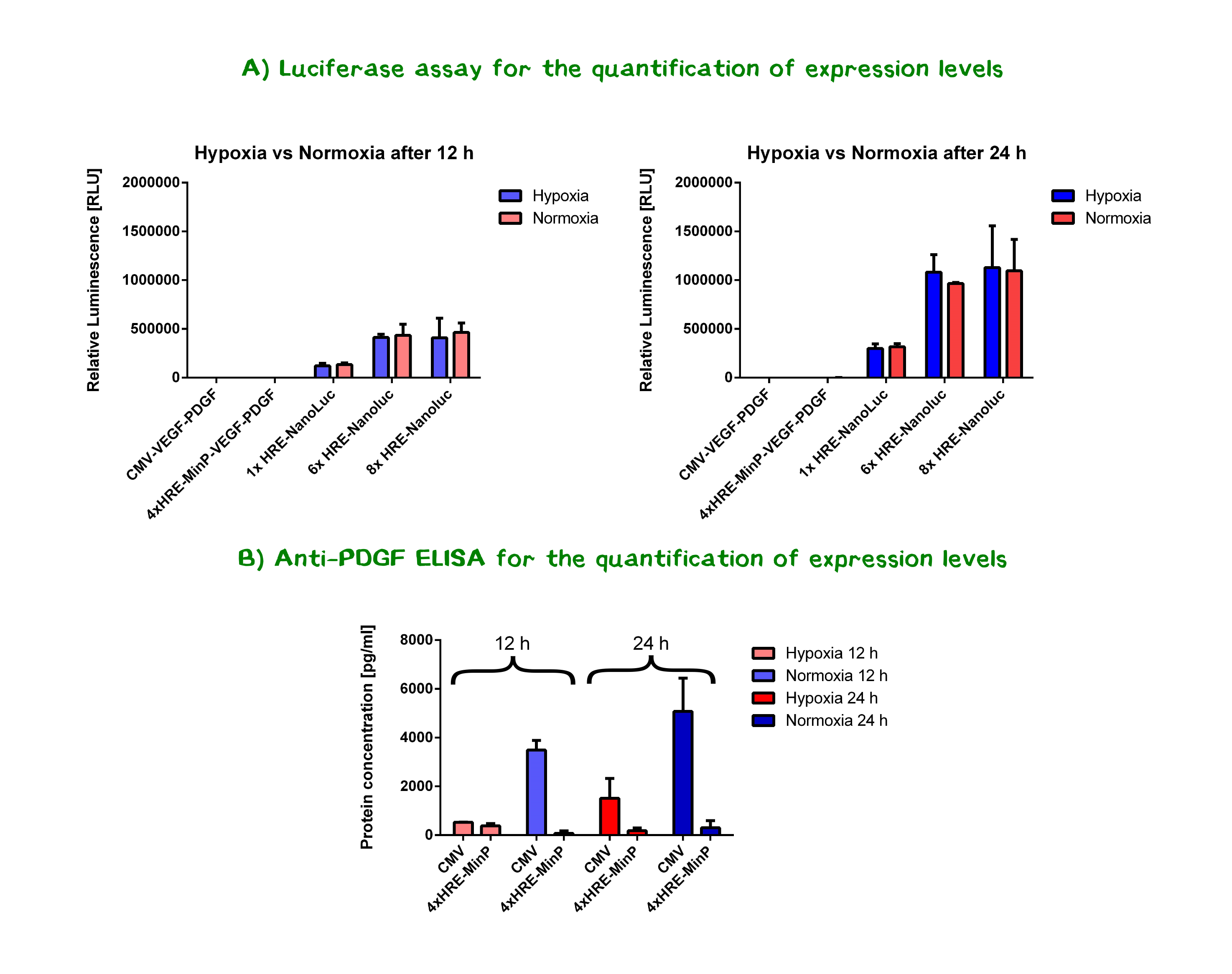
| + | [[File:Muc16 Hyp totale.png|thumb|center|830px|'''Figure 3:''' A) Results of the nanoluciferase assay for the quantification of expression levels. VEGF/PDGF-constructs act hereby as a negative control. Resulting luminescence was normalized by the respective luminescence value of the 0 h-sample. B) Results from the anti-PDGF ELISA measurements of secretion levels of PDGF protein in medium. Depicted are the protein concentrations in pg/ml as determined via a calibration line, that was calculated using PDGF protein standard samples.]] | ||
| − | |||
| − | |||
| − | |||
| − | |||
| − | |||
| − | |||
| − | |||
| − | |||
| − | |||
| − | |||
| + | '''Two interesting conclusions''' could be drawn from the results of the luciferase assays and the ELISA. | ||
| − | * | + | * A tendency could be observed for the luciferase constructs - showing that the '''number of HREs positively correlates with expression levels''' - the constructs with 6x and 8x HREs showed the highest expression/ secretion levels. In order to determine whether the differences between the nanoluciferase constructs containing 6x HRE and 8x HRE were statistically significant, a two-tailed t-test was performed. Resulting P-values of 0.98 and 0.87 for 12 h values, as well as 0.74 and 0.52 for 24 h (hypoxia and normoxia, respectively) and thus indicating that the results, by conventional definition, do not significantly differ. This may hint at the fact that '''more than 6 HREs do not increase promoter activity''' either in presence or abscence of oxygen. Further experiments may shed light on the matter. |
| − | * '''Expression levels compared between hypoxia and normoxia do ''not'' significantly differ''' at both time points , | + | * '''Expression levels compared between hypoxia and normoxia do ''not'' significantly differ''' at both time points , meaning the designed promoter does not significantly increase expression levels under the tested hypoxic conditions. Yet, considering that both transcription and translation are negatively influenced by hypoxic stress <ref>Strzyz, P. (2016). Cancer biology: Hypoxia as an off switch for gene expression. Nature Reviews Molecular Cell Biology.</ref> <ref>Liu, L., Cash, T. P., Jones, R. G., Keith, B., Thompson, C. B., & Simon, M. C. (2006). Hypoxia-induced energy stress regulates mRNA translation and cell growth. Molecular cell, 21(4), 521-531.</ref> the hypoxia-response elements might actually be counter-acting these effects, maintaining expression levels. Especially compared to the significant drop in CMV-activity under hypoxia as shown by the ELISA, the 6x/ 8x HRE-MinP-constructs are remarkably steady in the abscence of oxygen. |
</div> | </div> | ||
Latest revision as of 20:32, 1 December 2016
Fighting hypoxia in printed tissue
Unicellular organisms such as bacteria possess the ability to reproduce very quickly when given the right conditions such as warmth, moisture and suitable nutrients,[1] and enter an inactive state when these are limited. On the contrary, most multicellular systems depend on a steady supply of oxygen and nutrients essential for their survival and metabolic stability.
Herein lies one of the main challenges of bioprinting approaches. When printing, the homogenous delivery of oxygen to three-dimensional cell-constructs is limited by diffusion gradients in tissue from the periphery towards the center [2], thus resulting in tissue hypoxia. [3]
In our bodies, this need for oxygen supply is met by blood vessels, which form extensive networks that nurture all tissues. As a result, capillaries grow and regress in healthy tissues according to functional demands. Thereby, no metabolically active tissue in the body is more than a few hundred micrometers from a blood capillary, which is formed by the process of angiogenesis. [4]
Sprouting angiogenesis, as implied by its name is characterized by sprouts composed of endothelial cells, which usually grow toward an angiogenic stimulus.

This driving stimulus is initiated in poorly perfused tissues when oxygen sensing mechanisms detect a level of hypoxia that demands the formation of new blood vessels to satisfy the metabolic requirements of parenchymal cells. Most types of parenchymal cells respond to a hypoxic environment by secreting a key proangiogenic growth factor called vascular endothelial growth factor (VEGF-A). [6]
Because the production of a normal vasculature is among others heavily dependent upon the concentration of VEGF-A in the tissue, we have constructed a model in order to measure the hypoxia dependent secretion of VEGF and PDGF (the so-called platelet-derived growth factor, which stimualtes cell division[7]). Thus, we may simulate the extreme conditions that are present when printing several layers of tissue and, by triggering sprouting angiogenesis, induce growth of blood vessels towards portions of tissues previously devoid of them.
Hypoxia-inducible promoters
Hypoxia refers to subnormal levels of oxygen in air, blood and tissue. Tissue hypoxia leads to cellular dysfunction and can ultimately lead to cell death. The ability of cells to adapt to periods of hypoxia is therefore important for their survival. [8] In this context, we wanted to find the most suitable way to compensate the poor oxygen conditions that are generated when printing tissue structures with a volume of more than a few mm3.
Following a previously published idea by Lee, J. Y., et al. "A novel chimeric promoter that is highly responsive to hypoxia and metals." to enhance the level of hypoxic induction and to control target gene expression [9], we have designed a potent hypoxia-inducible promoter system, making use of so-called hypoxia-response elements.
Many hypoxia-responsive genes are regulated by the hypoxia-response element (HRE) protein family of transcription factors, including hypoxia-inducible factor-1 (HIF-1). [10] When HREs derived from different genes are placed in plasmids, they confer hypoxia inducibility upon heterologous promoters in various cell types. [11] We intend to use this system of hypoxia-inducible expression to induce vascularization in printed tissues.
Constructs and methodology
In order to test the responsivity of HREs, we incorporated different copy numbers (1x-HRE, 6x-HRE, 8x-HRE) upstream from a minimal promoter. The evaluation of the constructs' inductivity was performed by measuring the secretion of a luciferase reporter after transient transfection in HEK293T cells.
Parallely we expressed a HRE´s activated construct containing the actual growth factors VEGF and PDGF under the same conditions and compared it to a positive control consisting of a CMV driven VEGF and PDGF gene.
For the experiments cells were subjected to normoxia (21% O2, 5% CO2, 74% N2) and to hypoxia (1% O2, 7% CO2, 92% N2) for 24 hours.
By taking samples of the medium after 0 h, 12 h and 24 h after the exposure of the cells to this conditions and measuring luciferase activity by a corresponding assay, we were able to quantify hypoxia-dependent gene expression. To be able to determine how far gene expression is responsive to hypoxia in comparison to normal oxygen concentrations, a control at normoxic conditions was furthermore run.
For the assay, a total of five constructs were tested, three of which were luciferase constructs for the quantification of expression levels and two of which were VEGF-constructs that aimed to confirm the expression of the actual growth factor by our cells via ELISA, as well as acting as a negative control for the luciferase assay. The luciferase constructs all contained the hypoxia promoter.
Results

Two interesting conclusions could be drawn from the results of the luciferase assays and the ELISA.
- A tendency could be observed for the luciferase constructs - showing that the number of HREs positively correlates with expression levels - the constructs with 6x and 8x HREs showed the highest expression/ secretion levels. In order to determine whether the differences between the nanoluciferase constructs containing 6x HRE and 8x HRE were statistically significant, a two-tailed t-test was performed. Resulting P-values of 0.98 and 0.87 for 12 h values, as well as 0.74 and 0.52 for 24 h (hypoxia and normoxia, respectively) and thus indicating that the results, by conventional definition, do not significantly differ. This may hint at the fact that more than 6 HREs do not increase promoter activity either in presence or abscence of oxygen. Further experiments may shed light on the matter.
- Expression levels compared between hypoxia and normoxia do not significantly differ at both time points , meaning the designed promoter does not significantly increase expression levels under the tested hypoxic conditions. Yet, considering that both transcription and translation are negatively influenced by hypoxic stress [12] [13] the hypoxia-response elements might actually be counter-acting these effects, maintaining expression levels. Especially compared to the significant drop in CMV-activity under hypoxia as shown by the ELISA, the 6x/ 8x HRE-MinP-constructs are remarkably steady in the abscence of oxygen.
References
- ↑ http://www.bbc.co.uk/schools/gcsebitesize/science/ triple_ocr_gateway/beyond_the_microscope/understanding_microbes/revision/2/
- ↑ Volkmer, Elias, et al. "Hypoxia in static and dynamic 3D culture systems for tissue engineering of bone." Tissue Engineering Part A 14.8 (2008): 1331-1340
- ↑ Höckel, Michael, and Peter Vaupel. "Tumor hypoxia: definitions and current clinical, biologic, and molecular aspects." Journal of the National Cancer Institute 93.4 (2001): 266-276.
- ↑ Adair TH, Montani JP. Angiogenesis. San Rafael (CA): Morgan & Claypool Life Sciences; 2010. Chapter 1, Overview of Angiogenesis doi:10.1089/ten.tea.2007.0231
- ↑ Modified from: http://www.liquidarea.com/2011/05/al-cnr-arrivano-i-farmaci-intelligenti-terapie-del-futuro/
- ↑ Adair TH, Montani JP. Angiogenesis. San Rafael (CA): Morgan & Claypool Life Sciences; 2010. Chapter 1, Overview of Angiogenesis doi:10.1089/ten.tea.2007.0231
- ↑ Li, X., & Eriksson, U. (2003). Novel PDGF family members: PDGF-C and PDGF-D. Cytokine & growth factor reviews, 14(2), 91-98.
- ↑ Muz, Barbara, et al. "Hypoxia. The role of hypoxia and HIF-dependent signalling events in rheumatoid arthritis." Arthritis research & therapy 11.1 (2009): 1
- ↑ Lee, J. Y., et al. "A novel chimeric promoter that is highly responsive to hypoxia and metals." Gene therapy 13.10 (2006): 857-868
- ↑ Lee, J. Y., et al. "A novel chimeric promoter that is highly responsive to hypoxia and metals." Gene therapy 13.10 (2006): 857-868
- ↑ Boast K, Binley K, Iqball S, Price T, Spearman H, Kingsman S et al. Characterization of physiologically regulated vectors for the treatment of ischemic disease. Hum Gene Ther 1999; 10: 2197–2208
- ↑ Strzyz, P. (2016). Cancer biology: Hypoxia as an off switch for gene expression. Nature Reviews Molecular Cell Biology.
- ↑ Liu, L., Cash, T. P., Jones, R. G., Keith, B., Thompson, C. B., & Simon, M. C. (2006). Hypoxia-induced energy stress regulates mRNA translation and cell growth. Molecular cell, 21(4), 521-531.


