Proof
In our project, we have created an epinephrine sensing device combined the biobrick parts BBa_K1969007 and BBa_K1969019 together (Fig.1) which can be able to differentiate the epinephrine solution in a wide concentration change.
Nano-lantern(cAMP-1.6) regulated by ADH1 promoter (BBa_K1969007)
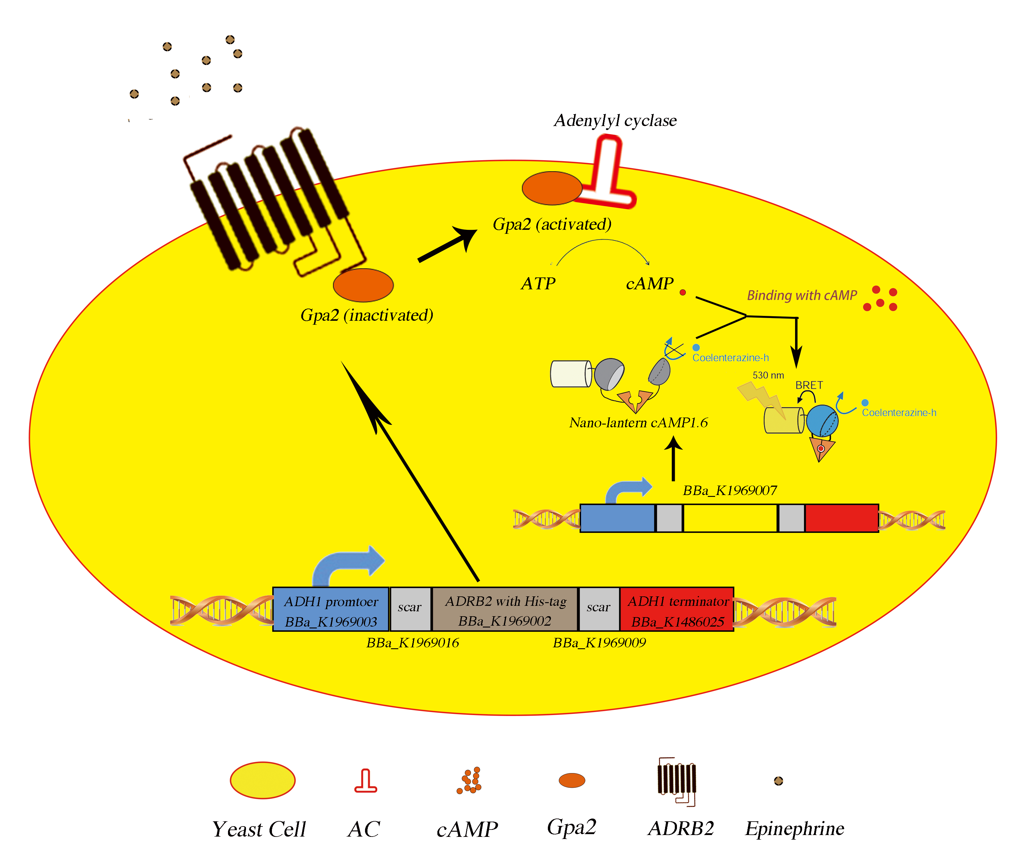
Fig.1 The picturized demonstration of working mechanisms in epinephrine sensing device (strain SMT-pANpAA)
We expressed the human adrenergic beta 2 downstream the ADH1 promoter and transformed into strain SMT-pAN to generate an epinephrine sensing strain, SMT-pANpAA. Actually, the cds for ADRB2 has already been registered as a biobrick part (BBa_K209472), while it’s not biobrick compatitble, so we use site mutation to make it a biobrick compatitble one (BBa_K1969001) and added a His-tag to its C terminus (BBa_K1969002).
We expressed the human adrenergic beta 2 downstream the ADH1 promoter and transformed into strain SMT-pAN to generate an epinephrine sensing strain, SMT-pANpAA. Actually, the cds for ADRB2 has already been registered as a biobrick part (BBa_K209472), while it’s not biobrick compatitble, so we use site mutation to make it a biobrick compatitble one (BBa_K1969001) and added a His-tag to its C terminus (BBa_K1969002).
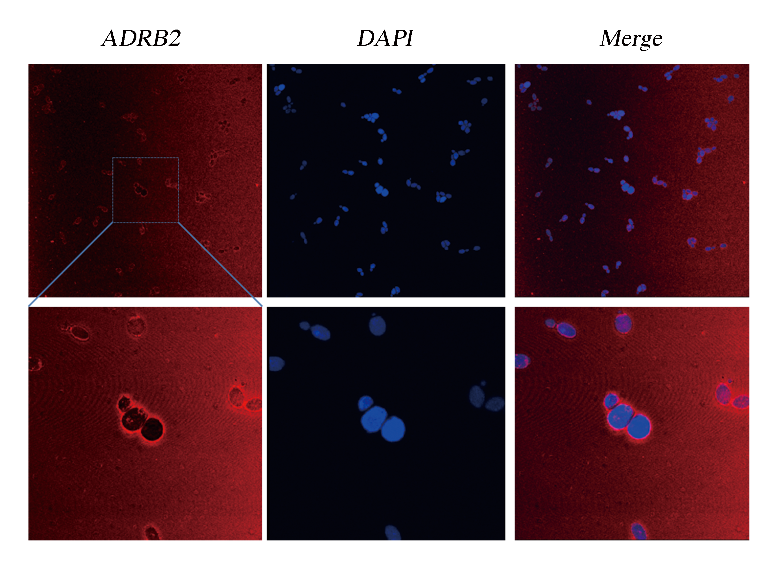
Fig.2 Detection of Membrane Localization of ADRB2 by Immunocytochemistry. The yeast cells were stabilized and stained with DAPI. As ADRB2 is expressed with a His-tag at C' terminal, thus anti-His mAb is used as the primary antibody and dunkey anti-mouse IgG 594 (Invitrogen) as the second antibody. Pictures were taken with Leica-SP5, 63 X.
Although the functional coupling of ADRB2 with yeast endogenous pheromone sensing pathway has been well studied in past few decades, the feasibility of functional signal transduction via ADRB2-Gpa2 has never been tested. So we used our engineered strains SMT-pANpAA as biosensors to detect extracellular epinephrine concentration in PBS solution. The response of the epinephrine is detected within first 5 minutes after addition of epinephrine (dissolved in HAc then diluted using PBS) (Fig.3).
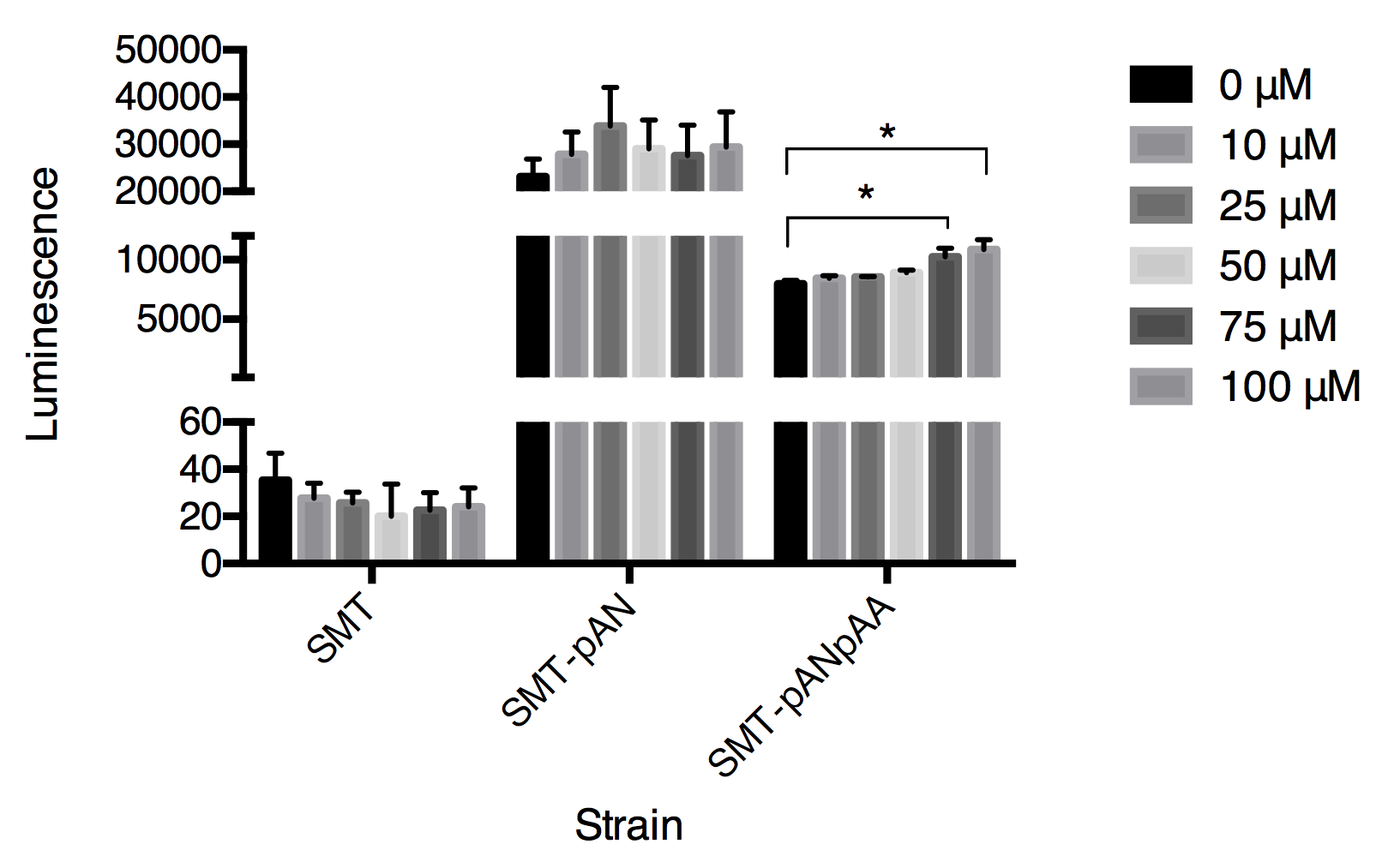
Fig.3 The luminescence triggered by epinephrine detected in three strains. Luminescence was collected at 30s after adding epinephrine. Data expressed as mean ± SD, significance was analyzed using Student-t test, n=3.
In this graph, we can tell that the constructed strain SMT-pANpAA was able to differentiate solution with relatively high concentration of epinephrine (75-100μM) with P value less than 0.05 by Student-t test. Although the difference between 100 μM and 75 μM treatment group was significant, the device can’t differentiate epinephrine solution at low concentration. And the expression of two exogenous protein under the same constitutive promoter ADH1 seems to interfere with each other as the overall luminescence decrease in strain SMT-pANpAA compared with strain SMT-pAN.
So far, we have succeeded in constructing an epinephrine sensor combining two composite parts, and the remaining questions will be further discussed in Vision For the Future part.
